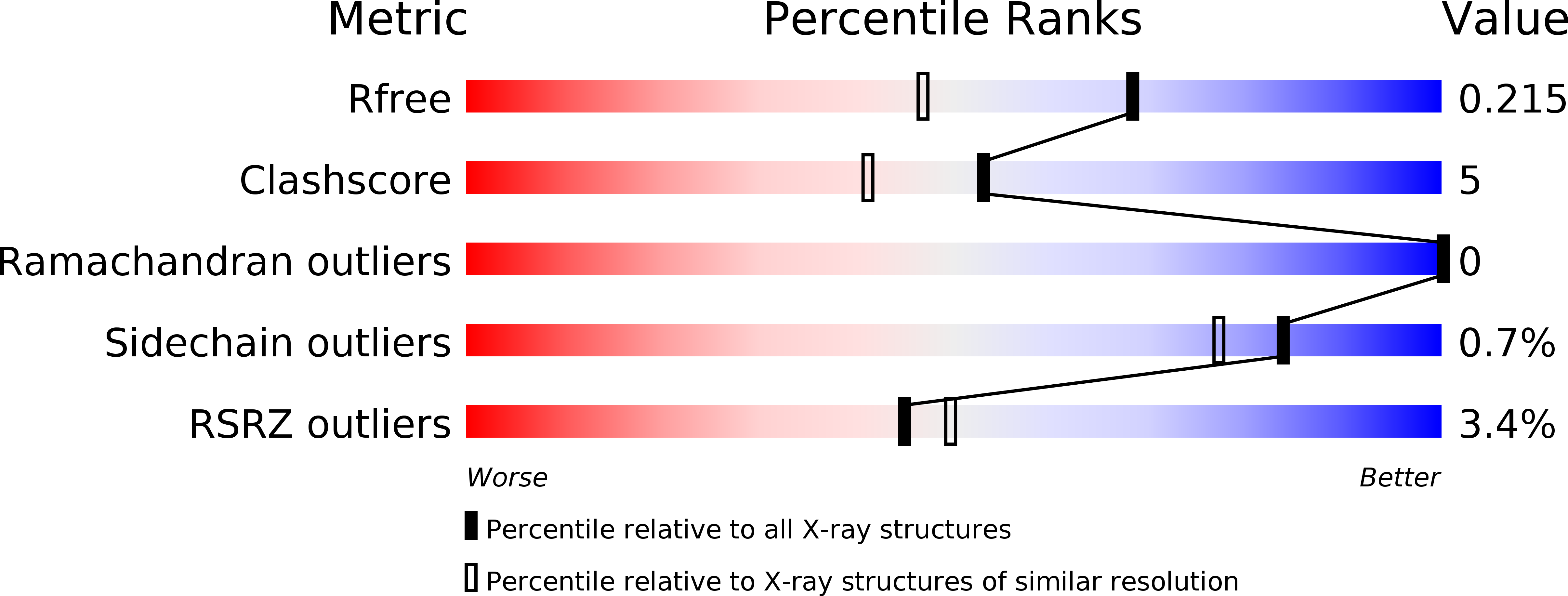
3BOD image
Deposition Date 2007-12-17
Release Date 2008-03-25
Last Version Date 2024-02-21
Entry Detail
PDB ID:
3BOD
Keywords:
Title:
Structure of mouse beta-neurexin 1
Biological Source:
Source Organism:
Mus musculus (Taxon ID: 10090)
Host Organism:
Method Details:
Experimental Method:
Resolution:
1.70 Å
R-Value Free:
0.22
R-Value Work:
0.17
R-Value Observed:
0.17
Space Group:
P 21 21 21
Macromolecular Entities
Structures with similar UniProt ID
Protein Blast
Polymer Type:polypeptide(L)
Molecule:Neurexin-1-alpha
Gene (Uniprot):Nrxn1
Chain IDs:A
Chain Length:178
Number of Molecules:1
Biological Source:Mus musculus
Ligand Molecules
Primary Citation
Crystal Structures of beta-Neurexin 1 and beta-Neurexin 2 Ectodomains and Dynamics of Splice Insertion Sequence 4.
Structure 16 410 421 (2008)
PMID: 18334216 DOI: 10.1016/j.str.2007.12.024

Abstact

Presynaptic neurexins (NRXs) bind to postsynaptic neuroligins (NLs) to form Ca(2+)-dependent complexes that bridge neural synapses. beta-NRXs bind NLs through their LNS domains, which contain a single site of alternative splicing (splice site 4) giving rise to two isoforms: +4 and Delta. We present crystal structures of the Delta isoforms of the LNS domains from beta-NRX1 and beta-NRX2, crystallized in the presence of Ca(2+) ions. The Ca(2+)-binding site is disordered in the beta-NRX2 structure, but the 1.7 A beta-NRX1 structure reveals a single Ca(2+) ion, approximately 12 A from the splice insertion site, with one coordinating ligand donated by a glutamic acid from an adjacent beta-NRX1 molecule. NMR studies of beta-NRX1+4 show that the insertion sequence is unstructured, and remains at least partially disordered in complex with NL. These results raise the possibility that beta-NRX insertion sequence 4 may function in roles independent of neuroligin binding.

Legend

Protein

Chemical

Disease

Primary Citation of related structures