3BM4 image
Deposition Date 2007-12-12
Release Date 2008-05-20
Last Version Date 2023-11-01
Entry Detail
PDB ID:
3BM4
Keywords:
Title:
Crystal Structure of Human ADP-ribose Pyrophosphatase NUDT5 In complex with magnesium and AMPcpr
Biological Source:
Source Organism:
Homo sapiens (Taxon ID: 9606)
Host Organism:
Method Details:
Experimental Method:
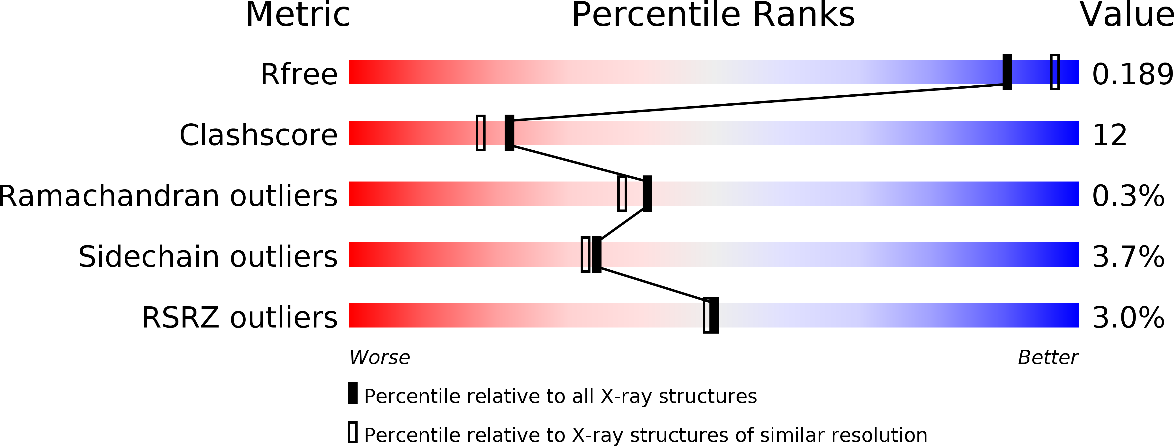
Resolution:
2.00 Å
R-Value Free:
0.23
R-Value Work:
0.19
R-Value Observed:
0.19
Space Group:
C 1 2 1
Macromolecular Entities
Polymer Type:polypeptide(L)
Molecule:ADP-sugar pyrophosphatase
Gene (Uniprot):NUDT5
Chain IDs:A, B
Chain Length:210
Number of Molecules:2
Biological Source:Homo sapiens
Primary Citation
Molecular Mechanism of ADP-Ribose Hydrolysis By Human NUDT5 From Structural and Kinetic Studies
J.Mol.Biol. 379 568 578 (2008)
PMID: 18462755 DOI: 10.1016/j.jmb.2008.04.006

Abstact

Human NUDT5 (hNUDT5) is an ADP-ribose (ADPR) pyrophosphatase (ADPRase) that plays important roles in controlling the intracellular levels of ADPR and preventing non-enzymatic ADP-ribosylation of proteins by hydrolyzing ADPR to AMP and ribose 5'-phosphate. We report the crystal structure of hNUDT5 in complex with a non-hydrolyzable ADPR analogue, alpha,beta-methyleneadenosine diphosphoribose, and three Mg(2+) ions representing the transition state of the enzyme during catalysis. Analysis of this structure and comparison with previously reported hNUDT5 structures identify key residues involved in substrate binding and catalysis. In the transition-state structure, three metal ions are bound at the active site and are coordinated by surrounding residues and water molecules. A conserved water molecule is at an ideal position for nucleophilic attack on the alpha-phosphate of ADPR. The side chain of Glu166 on loop L9 changes its conformation to interact with the conserved water molecule compared with that in the substrate-bound structure and appears to function as a catalytic base. Mutagenesis and kinetic studies show that Trp28 and Trp46 are important for the substrate binding; Arg51 is involved in both the substrate binding and the catalysis; and Glu112 and Glu116 of the Nudix motif, Glu166 on loop L9, and Arg111 are critical for the catalysis. The structural and biochemical data together reveal the molecular basis of the catalytic mechanism of ADPR hydrolysis by hNUDT5. Specifically, Glu166 functions as a catalytic base to deprotonate a conserved water molecule that acts as a nucleophile to attack the alpha-phosphate of ADPR, and three Mg(2+) ions are involved in the activation of the nucleophile and the binding of the substrate. Structural comparison of different ADPRases also suggests that most dimeric ADPRases may share a similar catalytic mechanism of ADPR hydrolysis.

Legend

Protein

Chemical

Disease

Primary Citation of related structures