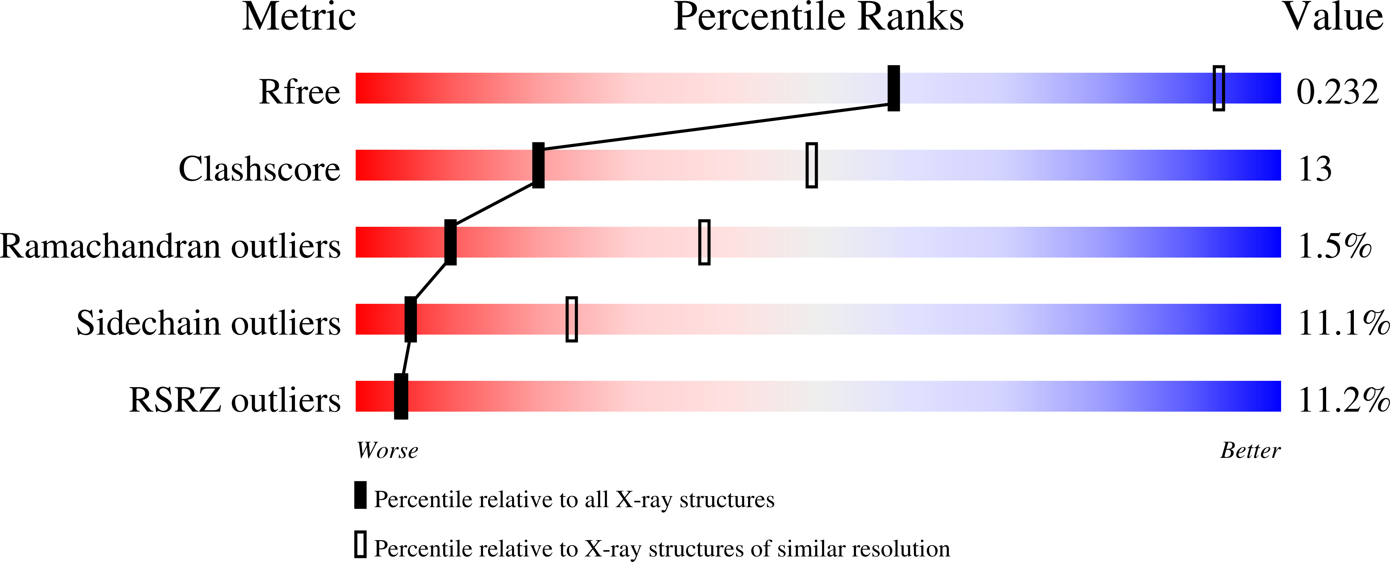
3BL8 image
Deposition Date 2007-12-10
Release Date 2008-02-19
Last Version Date 2024-10-30
Entry Detail
PDB ID:
3BL8
Keywords:
Title:
Crystal structure of the extracellular domain of neuroligin 2A from mouse
Biological Source:
Source Organism:
Mus musculus (Taxon ID: 10090)
Host Organism:
Method Details:
Experimental Method:
Resolution:
3.30 Å
R-Value Free:
0.26
R-Value Work:
0.21
R-Value Observed:
0.22
Space Group:
C 1 2 1
Macromolecular Entities
Structures with similar UniProt ID
Protein Blast
Polymer Type:polypeptide(L)
Molecule:Neuroligin-2
Gene (Uniprot):Nlgn2
Chain IDs:A, B, C, D
Chain Length:580
Number of Molecules:4
Biological Source:Mus musculus
Modified Residue
Compound ID Chain ID Parent Comp ID Details 2D Image
ASN A ASN GLYCOSYLATION SITE
Ligand Molecules
Primary Citation
Crystal structure of the extracellular cholinesterase-like domain from neuroligin-2.
Proc.Natl.Acad.Sci.Usa 105 1873 1878 (2008)
PMID: 18250328 DOI: 10.1073/pnas.0711701105

Abstact

Neuroligins (NLs) are catalytically inactive members of a family of cholinesterase-like transmembrane proteins that mediate cell adhesion at neuronal synapses. Postsynaptic neuroligins engage in Ca2+-dependent transsynaptic interactions via their extracellular cholinesterase domain with presynaptic neurexins (NRXs). These interactions may be regulated by two short splice insertions (termed A and B) in the NL cholinesterase domain. Here, we present the 3.3-A crystal structure of the ectodomain from NL2 containing splice insertion A (NL2A). The overall structure of NL2A resembles that of cholinesterases, but several structural features are unique to the NL proteins. First, structural elements surrounding the esterase active-site region differ significantly between active esterases and NL2A. On the opposite surface of the NL2A molecule, the positions of the A and B splice insertions identify a candidate NRX interaction site of the NL protein. Finally, sequence comparisons of NL isoforms allow for mapping the location of residues of previously identified mutations in NL3 and NL4 found in patients with autism spectrum disorders. Overall, the NL2 structure promises to provide a valuable model for dissecting NL isoform- and synapse-specific functions.

Legend

Protein

Chemical

Disease

Primary Citation of related structures