Deposition Date
2007-12-06
Release Date
2008-01-29
Last Version Date
2024-11-20
Entry Detail
PDB ID:
3BKD
Keywords:
Title:
High resolution Crystal structure of Transmembrane domain of M2 protein
Method Details:
Experimental Method:
Resolution:
2.05 Å
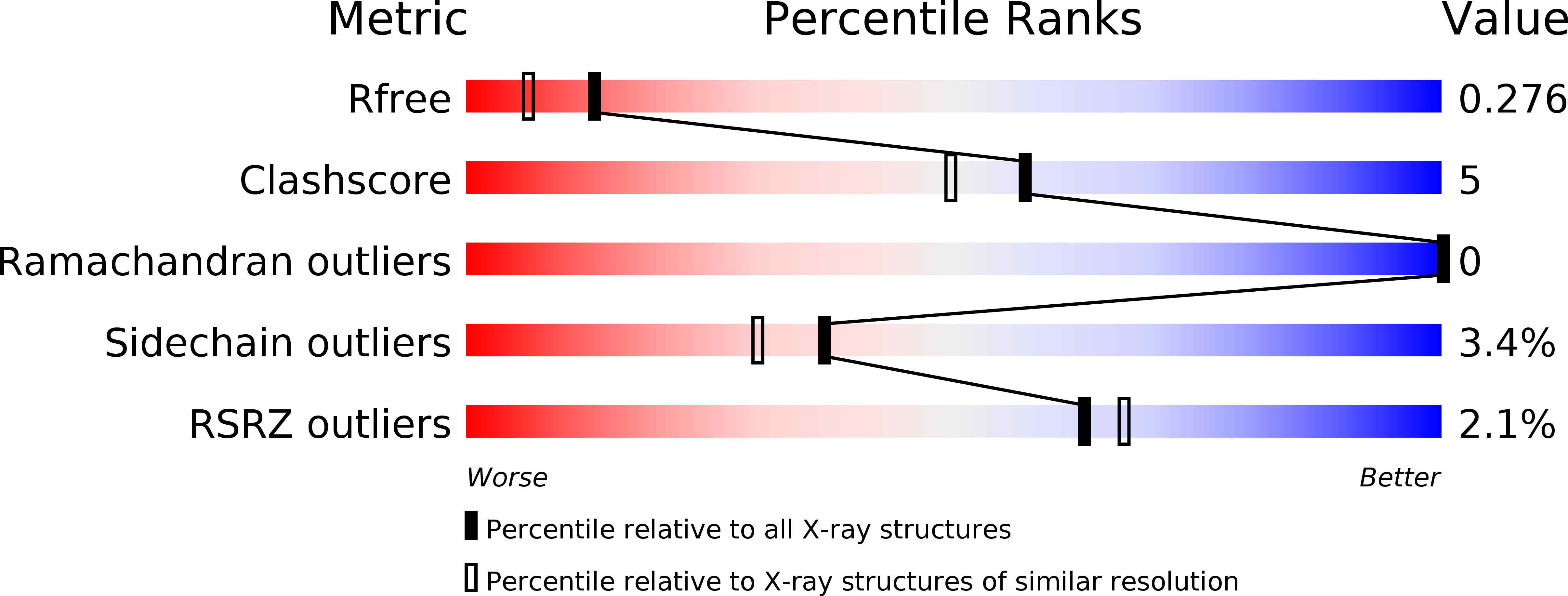
R-Value Free:
0.26
R-Value Work:
0.21
R-Value Observed:
0.22
Space Group:
P 1 21 1