Deposition Date
2007-11-30
Release Date
2008-12-09
Last Version Date
2023-08-30
Entry Detail
PDB ID:
3BIG
Keywords:
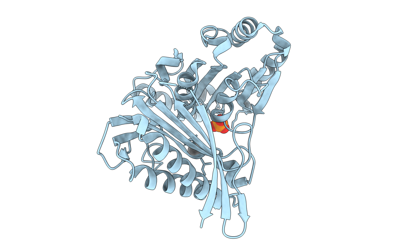
Title:
Crystal structure of the fructose-1,6-bisphosphatase GlpX from E.coli in complex with inorganic phosphate
Biological Source:
Source Organism(s):
Escherichia coli (Taxon ID: )
Expression System(s):
Method Details:
Experimental Method:
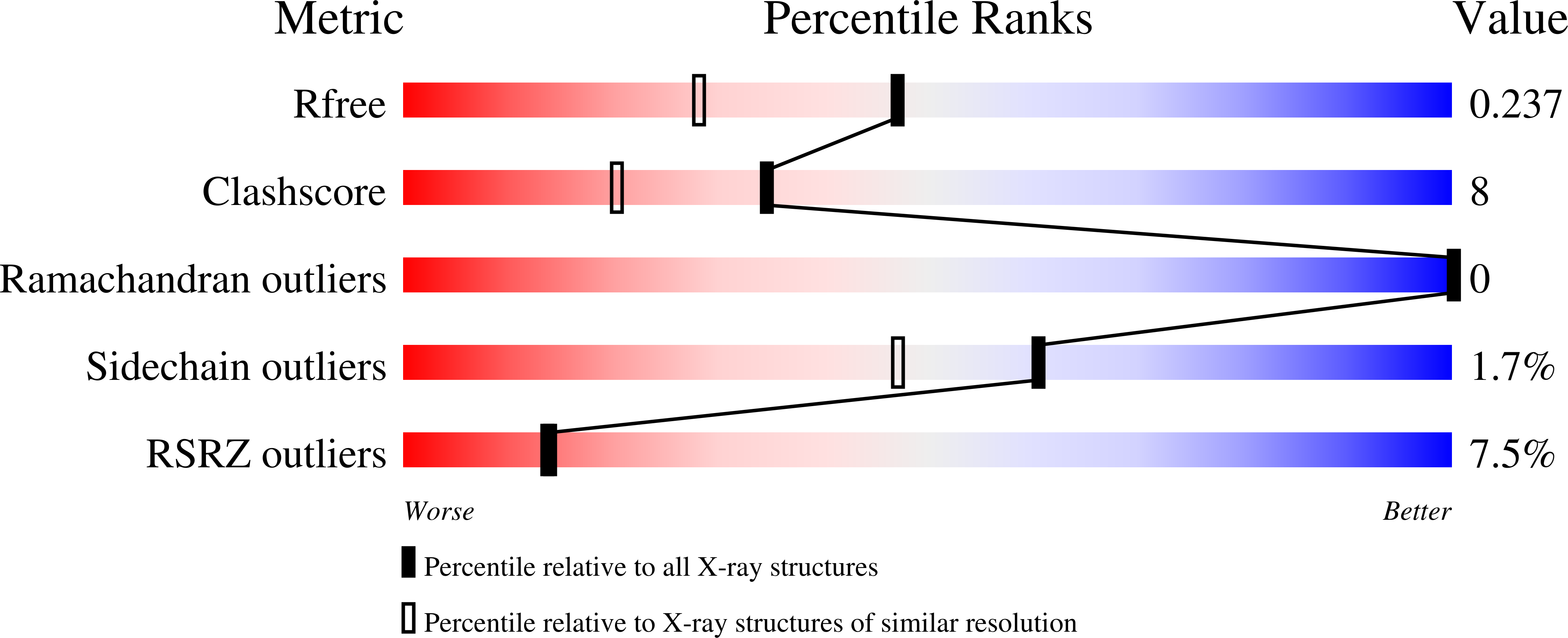
Resolution:
1.85 Å
R-Value Free:
0.23
R-Value Work:
0.18
R-Value Observed:
0.19
Space Group:
P 4 2 2