3BFP image
Deposition Date 2007-11-22
Release Date 2008-01-22
Last Version Date 2024-02-21
Entry Detail
PDB ID:
3BFP
Keywords:
Title:
Crystal Structure of apo-PglD from Campylobacter jejuni
Biological Source:
Source Organism:
Host Organism:
Method Details:
Experimental Method:
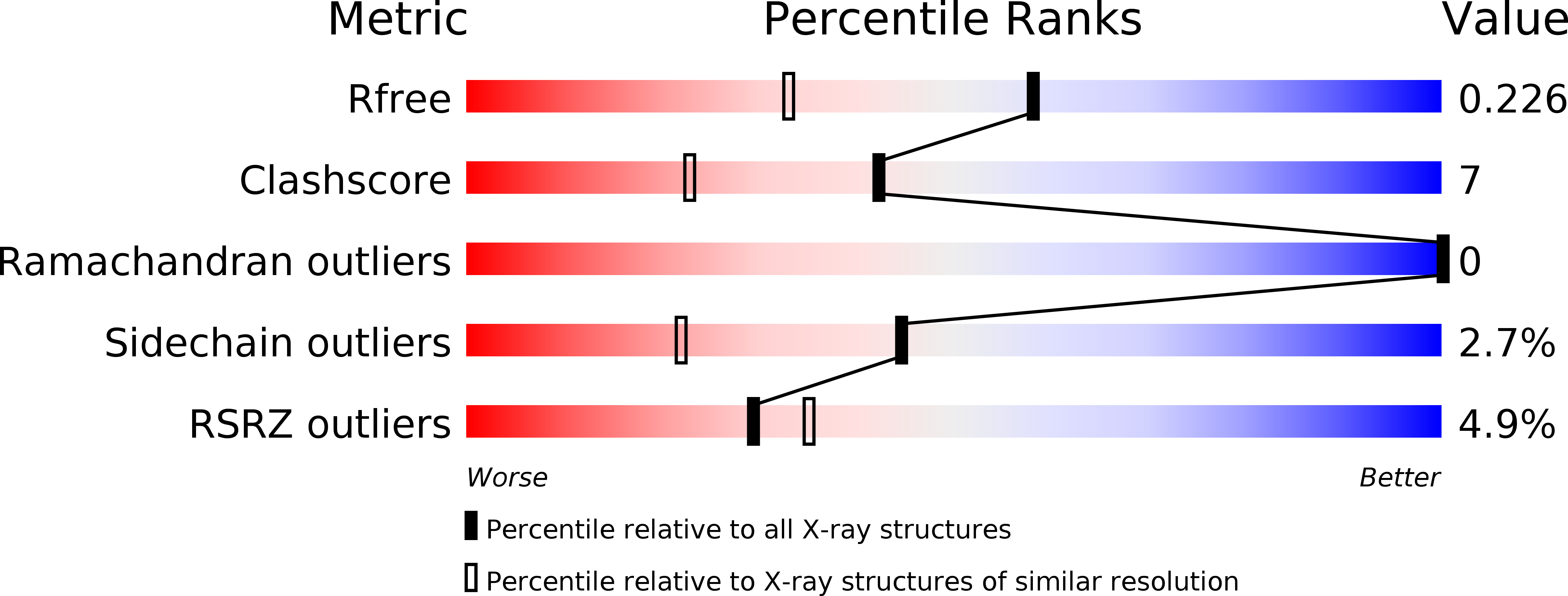
Resolution:
1.75 Å
R-Value Free:
0.22
R-Value Work:
0.19
R-Value Observed:
0.20
Space Group:
P 63
Macromolecular Entities
Polymer Type:polypeptide(L)
Molecule:Acetyltransferase
Gene (Uniprot):pglD
Chain IDs:A
Chain Length:194
Number of Molecules:1
Biological Source:Campylobacter jejuni
Ligand Molecules
Primary Citation
Structure and Active Site Residues of PglD, an N-Acetyltransferase from the Bacillosamine Synthetic Pathway Required for N-Glycan Synthesis in Campylobacter jejuni.
Biochemistry 47 1827 1836 (2008)
PMID: 18198901 DOI: 10.1021/bi702032r

Abstact

Campylobacter jejuni is highly unusual among bacteria in forming N-linked glycoproteins. The heptasaccharide produced by its pgl system is attached to protein Asn through its terminal 2,4-diacetamido-2,4,6-trideoxy-d-Glc (QuiNAc4NAc or N,N'-diacetylbacillosamine) moiety. The crucial, last part of this sugar's synthesis is the acetylation of UDP-2-acetamido-4-amino-2,4,6-trideoxy-d-Glc by the enzyme PglD, with acetyl-CoA as a cosubstrate. We have determined the crystal structures of PglD in CoA-bound and unbound forms, refined to 1.8 and 1.75 A resolution, respectively. PglD is a trimer of subunits each comprised of two domains, an N-terminal alpha/beta-domain and a C-terminal left-handed beta-helix. Few structural differences accompany CoA binding, except in the C-terminal region following the beta-helix (residues 189-195), which adopts an extended structure in the unbound form and folds to extend the beta-helix upon binding CoA. Computational molecular docking suggests a different mode of nucleotide-sugar binding with respect to the acetyl-CoA donor, with the molecules arranged in an "L-shape", compared with the "in-line" orientation in related enzymes. Modeling indicates that the oxyanion intermediate would be stabilized by the NH group of Gly143', with His125' the most likely residue to function as a general base, removing H+ from the amino group prior to nucleophilic attack at the carbonyl carbon of acetyl-CoA. Site-specific mutations of active site residues confirmed the importance of His125', Glu124', and Asn118. We conclude that Asn118 exerts its function by stabilizing the intricate hydrogen bonding network within the active site and that Glu124' may function to increase the pKa of the putative general base, His125'.

Legend

Protein

Chemical

Disease

Primary Citation of related structures