3BEO image
Deposition Date 2007-11-19
Release Date 2008-02-19
Last Version Date 2024-02-21
Entry Detail
PDB ID:
3BEO
Keywords:
Title:
A Structural Basis for the allosteric regulation of non-hydrolyzing UDP-GlcNAc 2-epimerases
Biological Source:
Source Organism:
Host Organism:
Method Details:
Experimental Method:
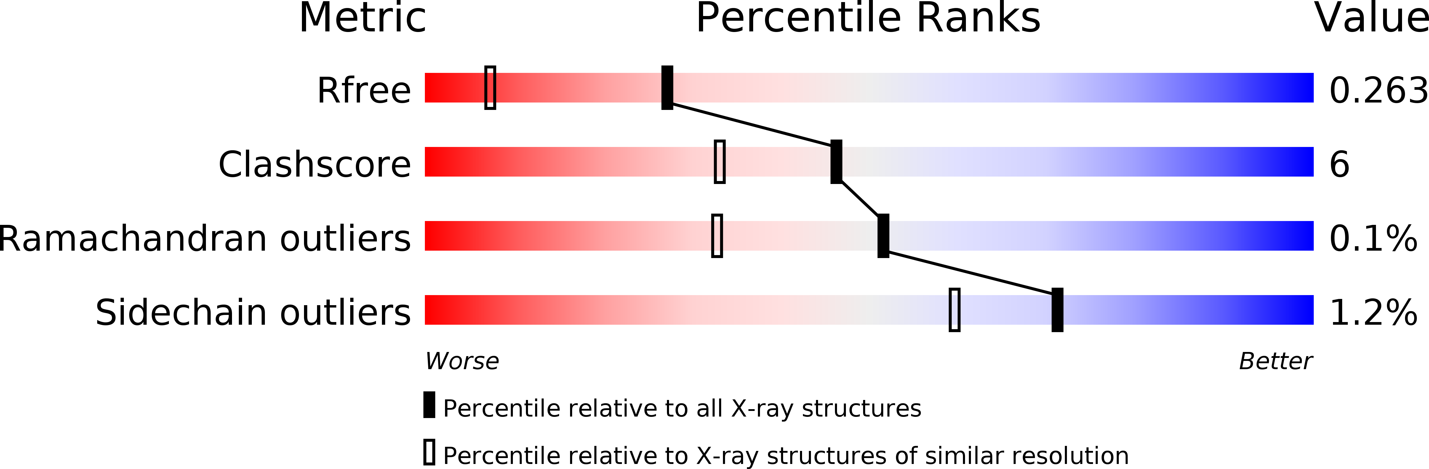
Resolution:
1.70 Å
R-Value Free:
0.26
R-Value Work:
0.22
R-Value Observed:
0.22
Space Group:
P 1 21 1
Macromolecular Entities
Structures with similar UniProt ID
Protein Blast
Polymer Type:polypeptide(L)
Molecule:UDP-N-acetylglucosamine 2-epimerase
Chain IDs:A, B
Chain Length:375
Number of Molecules:2
Biological Source:Bacillus anthracis
Primary Citation
A structural basis for the allosteric regulation of non-hydrolysing UDP-GlcNAc 2-epimerases.
Embo Rep. 9 199 205 (2008)
PMID: 18188181 DOI: 10.1038/sj.embor.7401154

Abstact

The non-hydrolysing bacterial UDP-N-acetylglucosamine 2-epimerase (UDP-GlcNAc 2-epimerase) catalyses the conversion of UDP-GlcNAc into UDP-N-acetylmannosamine, an intermediate in the biosynthesis of several cell-surface polysaccharides. This enzyme is allosterically regulated by its substrate UDP-GlcNAc. The structure of the ternary complex between the Bacillus anthracis UDP-GlcNAc 2-epimerase, its substrate UDP-GlcNAc and the reaction intermediate UDP, showed direct interactions between UDP and its substrate, and between the complex and highly conserved enzyme residues, identifying the allosteric site of the enzyme. The binding of UDP-GlcNAc is associated with conformational changes in the active site of the enzyme. Kinetic data and mutagenesis of the highly conserved UDP-GlcNAc-interacting residues confirm their importance in the substrate binding and catalysis of the enzyme. This constitutes the first example to our knowledge, of an enzymatic allosteric activation by direct interaction between the substrate and the allosteric activator.

Legend

Protein

Chemical

Disease

Primary Citation of related structures
Feedback Form
Name
Email
Institute
Feedback