3BEF image
Deposition Date 2007-11-17
Release Date 2008-01-01
Last Version Date 2024-10-30
Entry Detail
PDB ID:
3BEF
Keywords:
Title:
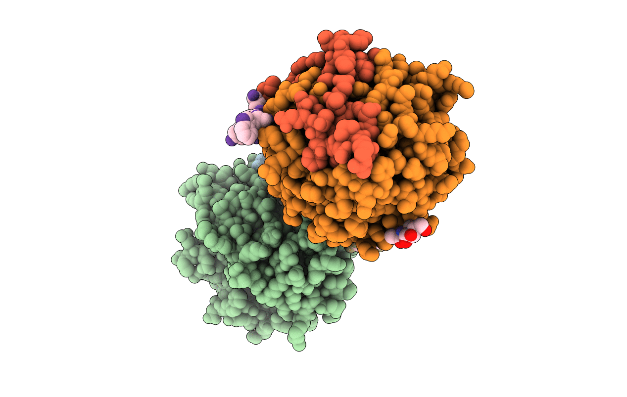
Crystal structure of thrombin bound to the extracellular fragment of PAR1
Biological Source:
Source Organism:
Homo sapiens (Taxon ID: 9606)
Host Organism:
Method Details:
Experimental Method:
Resolution:
2.20 Å
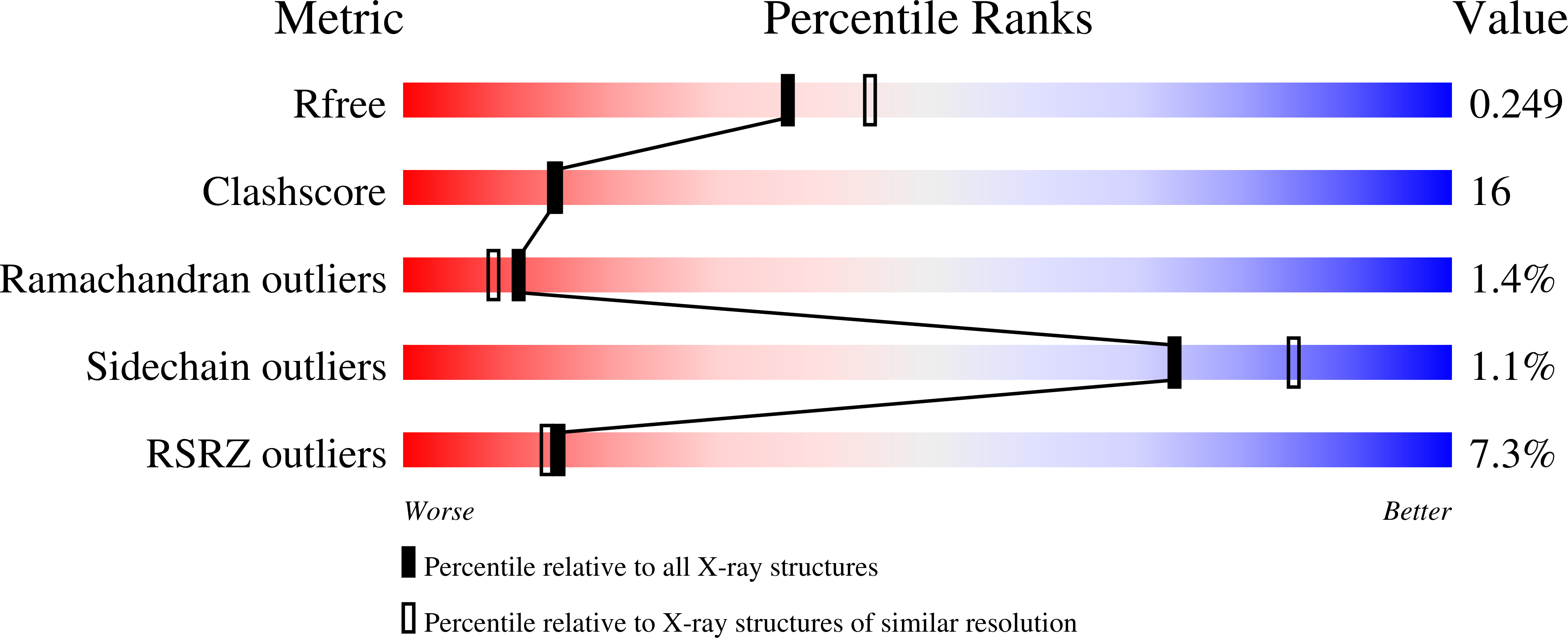
R-Value Free:
0.24
R-Value Work:
0.20
R-Value Observed:
0.20
Space Group:
P 1
Macromolecular Entities
Structures with similar UniProt ID
Protein Blast
Polymer Type:polypeptide(L)
Molecule:Prothrombin
Gene (Uniprot):F2
Mutagens:D102N
Chain IDs:A, D
Chain Length:46
Number of Molecules:2
Biological Source:Homo sapiens
Structures with similar UniProt ID
Protein Blast
Polymer Type:polypeptide(L)
Molecule:Prothrombin
Gene (Uniprot):F2
Chain IDs:B, E
Chain Length:259
Number of Molecules:2
Biological Source:Homo sapiens
Structures with similar UniProt ID
Protein Blast
Polymer Type:polypeptide(L)
Molecule:Proteinase-activated receptor 1
Gene (Uniprot):F2R
Chain IDs:C, F
Chain Length:9
Number of Molecules:2
Biological Source:Homo sapiens
Modified Residue
Compound ID Chain ID Parent Comp ID Details 2D Image
ASN B ASN GLYCOSYLATION SITE
Ligand Molecules
Primary Citation
Structural identification of the pathway of long-range communication in an allosteric enzyme.
Proc.Natl.Acad.Sci.Usa 105 1832 1837 (2008)
PMID: 18250335 DOI: 10.1073/pnas.0710894105

Abstact

Allostery is a common mechanism of regulation of enzyme activity and specificity, and its signatures are readily identified from functional studies. For many allosteric systems, structural evidence exists of long-range communication among protein domains, but rarely has this communication been traced to a detailed pathway. The thrombin mutant D102N is stabilized in a self-inhibited conformation where access to the active site is occluded by a collapse of the entire 215-219 beta-strand. Binding of a fragment of the protease activated receptor PAR1 to exosite I, 30-A away from the active site region, causes a large conformational change that corrects the position of the 215-219 beta-strand and restores access to the active site. The crystal structure of the thrombin-PAR1 complex, solved at 2.2-A resolution, reveals the details of this long-range allosteric communication in terms of a network of polar interactions.

Legend

Protein

Chemical

Disease

Primary Citation of related structures