3BDN image
Deposition Date 2007-11-15
Release Date 2008-04-15
Last Version Date 2023-08-30
Entry Detail
PDB ID:
3BDN
Title:
Crystal Structure of the Lambda Repressor
Biological Source:
Source Organism:
Method Details:
Experimental Method:
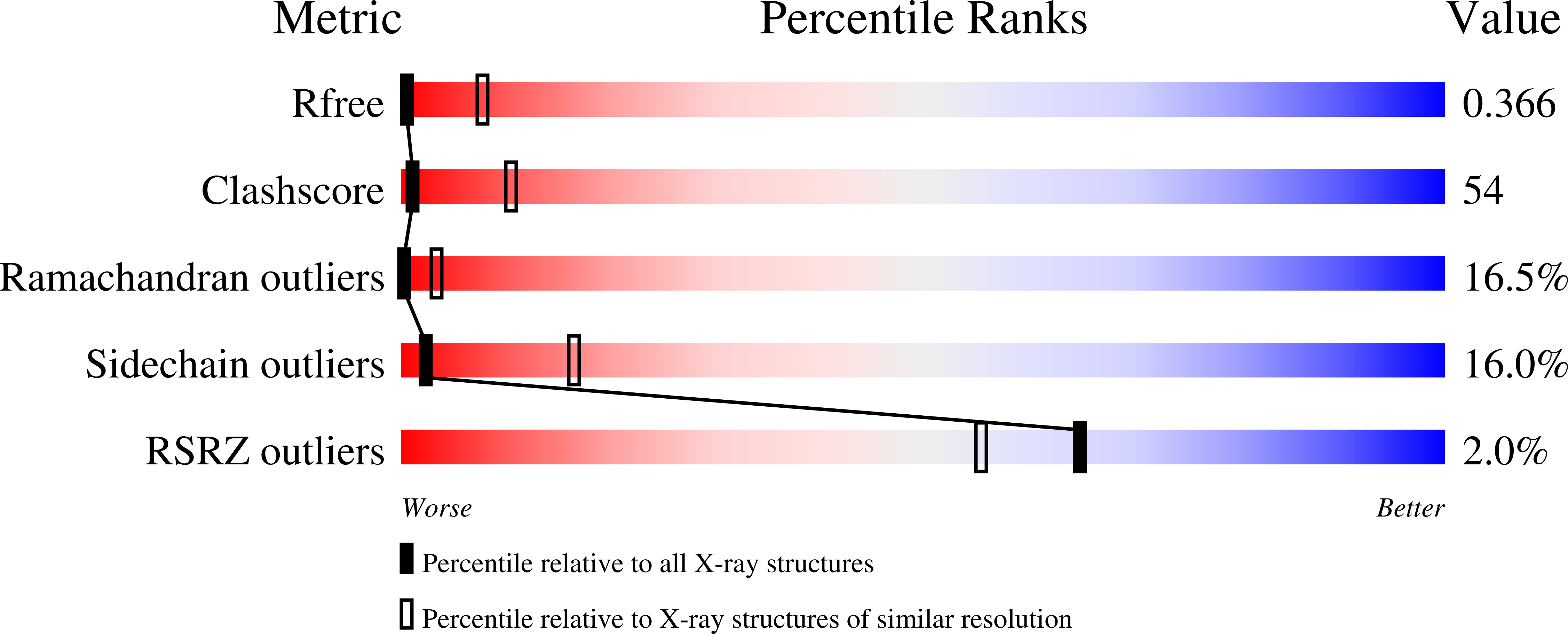
Resolution:
3.91 Å
R-Value Free:
0.37
R-Value Work:
0.29
R-Value Observed:
0.29
Space Group:
C 2 2 2
Macromolecular Entities
Structures with similar UniProt ID
Protein Blast
Polymer Type:polypeptide(L)
Molecule:Lambda Repressor
Gene (Uniprot):cI
Mutagens:D197G
Chain IDs:C (auth: A), D (auth: B)
Chain Length:236
Number of Molecules:2
Biological Source:Enterobacteria phage lambda
Ligand Molecules
Primary Citation
Crystal structure of the lambda repressor and a model for pairwise cooperative operator binding
Nature 452 1022 1025 (2008)
PMID: 18432246 DOI: 10.1038/nature06831

Abstact

Bacteriophage lambda has for many years been a model system for understanding mechanisms of gene regulation. A 'genetic switch' enables the phage to transition from lysogenic growth to lytic development when triggered by specific environmental conditions. The key component of the switch is the cI repressor, which binds to two sets of three operator sites on the lambda chromosome that are separated by about 2,400 base pairs (bp). A hallmark of the lambda system is the pairwise cooperativity of repressor binding. In the absence of detailed structural information, it has been difficult to understand fully how repressor molecules establish the cooperativity complex. Here we present the X-ray crystal structure of the intact lambda cI repressor dimer bound to a DNA operator site. The structure of the repressor, determined by multiple isomorphous replacement methods, reveals an unusual overall architecture that allows it to adopt a conformation that appears to facilitate pairwise cooperative binding to adjacent operator sites.

Legend

Protein

Chemical

Disease

Primary Citation of related structures
Feedback Form
Name
Email
Institute
Feedback