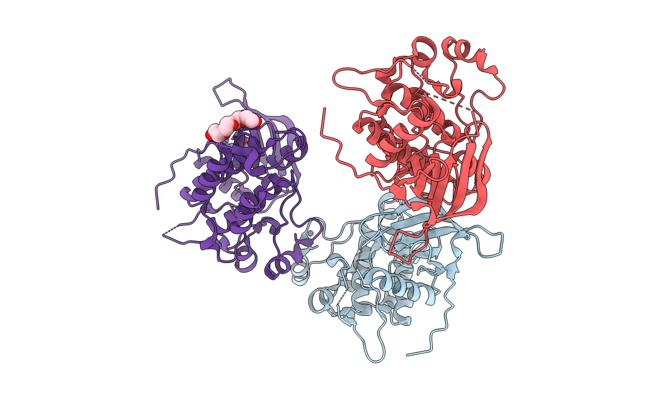
3BCE image
Deposition Date 2007-11-12
Release Date 2008-02-12
Last Version Date 2024-02-21
Entry Detail
PDB ID:
3BCE
Keywords:
Title:
Crystal structure of the ErbB4 kinase
Biological Source:
Source Organism:
Homo sapiens (Taxon ID: 9606)
Host Organism:
Method Details:
Experimental Method:
Resolution:
2.50 Å
R-Value Free:
0.25
R-Value Work:
0.19
R-Value Observed:
0.19
Space Group:
P 32
Macromolecular Entities
Polymer Type:polypeptide(L)
Molecule:Receptor tyrosine-protein kinase erbB-4
Gene (Uniprot):ERBB4
Chain IDs:A, B, C
Chain Length:328
Number of Molecules:3
Biological Source:Homo sapiens
Primary Citation
Mechanism of Activation and Inhibition of the HER4/ErbB4 Kinase.
Structure 16 460 467 (2008)
PMID: 18334220 DOI: 10.1016/j.str.2007.12.016

Abstact

HER4/ErbB4 is a ubiquitously expressed member of the EGF/ErbB family of receptor tyrosine kinases that is essential for normal development of the heart, nervous system, and mammary gland. We report here crystal structures of the ErbB4 kinase domain in active and lapatinib-inhibited forms. Active ErbB4 kinase adopts an asymmetric dimer conformation essentially identical to that observed to be important for activation of the EGF receptor/ErbB1 kinase. Mutagenesis studies of intact ErbB4 in Ba/F3 cells confirm the importance of this asymmetric dimer for activation of intact ErbB4. Lapatinib binds to an inactive form of the ErbB4 kinase in a mode equivalent to its interaction with the EGF receptor. All ErbB4 residues contacted by lapatinib are conserved in the EGF receptor and HER2/ErbB2, which lapatinib also targets. These results demonstrate that key elements of kinase activation and inhibition are conserved among ErbB family members.

Legend

Protein

Chemical

Disease

Primary Citation of related structures