3B9K image
Deposition Date 2007-11-05
Release Date 2008-11-11
Last Version Date 2024-10-30
Entry Detail
PDB ID:
3B9K
Keywords:
Title:
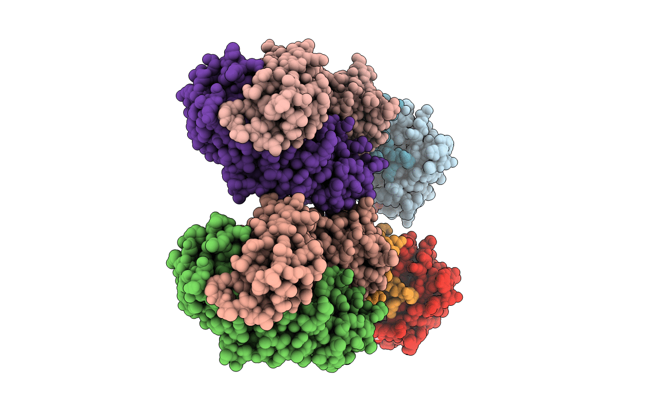
Crystal structure of CD8alpha-beta in complex with YTS 156.7 FAB
Biological Source:
Source Organism:
Mus musculus (Taxon ID: 10090)
rattus rattus (Taxon ID: 10117)
Method Details:
Experimental Method:
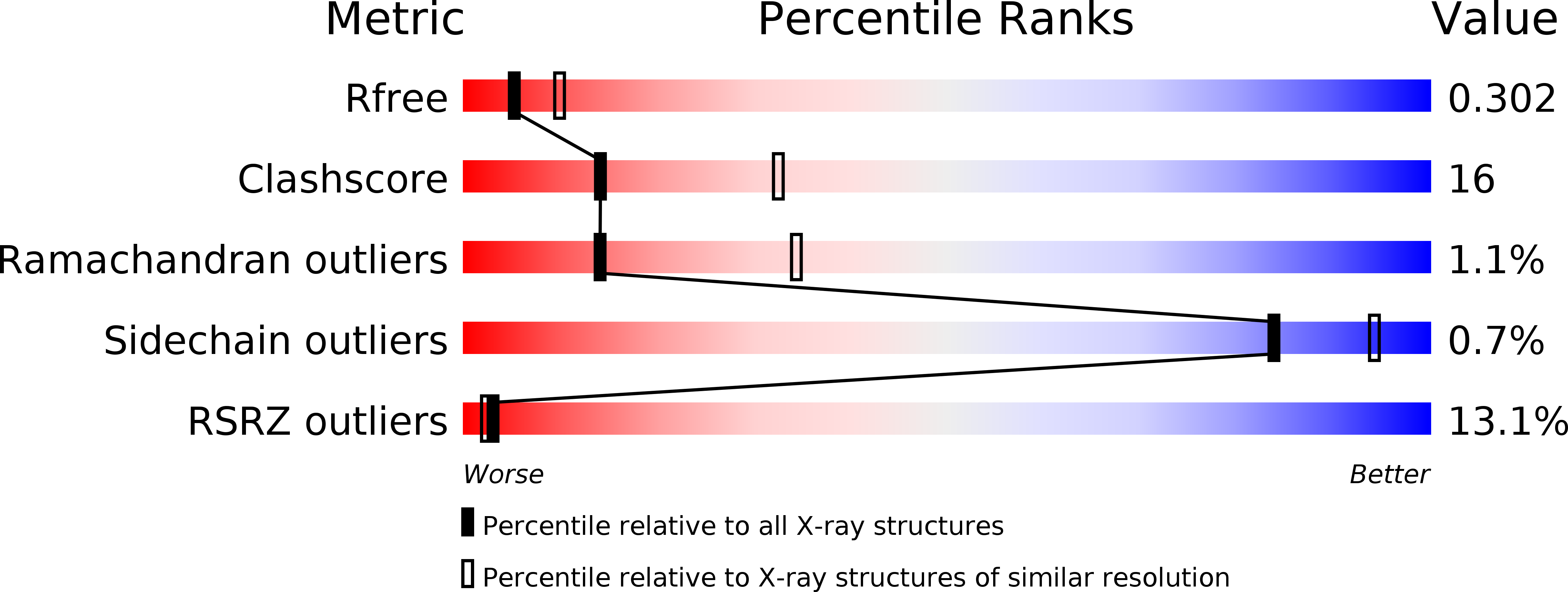
Resolution:
2.70 Å
R-Value Free:
0.28
R-Value Work:
0.23
R-Value Observed:
0.23
Space Group:
P 21 21 21
Macromolecular Entities
Structures with similar UniProt ID
Protein Blast
Polymer Type:polypeptide(L)
Molecule:T-cell surface glycoprotein CD8 alpha chain
Gene (Uniprot):Cd8a
Chain IDs:C (auth: A), G (auth: E)
Chain Length:131
Number of Molecules:2
Biological Source:Mus musculus
Structures with similar UniProt ID
Protein Blast
Polymer Type:polypeptide(L)
Molecule:T-cell surface glycoprotein CD8 beta chain
Gene (Uniprot):Cd8b
Chain IDs:D (auth: B), H (auth: F)
Chain Length:125
Number of Molecules:2
Biological Source:Mus musculus
Polymer Type:polypeptide(L)
Molecule:Fab Heavy chain
Chain IDs:B (auth: H), F (auth: D)
Chain Length:214
Number of Molecules:2
Biological Source:rattus rattus
Polymer Type:polypeptide(L)
Molecule:Fab Light Chain
Chain IDs:A (auth: L), E (auth: C)
Chain Length:213
Number of Molecules:2
Biological Source:rattus rattus
Modified Residue
Compound ID Chain ID Parent Comp ID Details 2D Image
ASN G ASN GLYCOSYLATION SITE
Ligand Molecules
Primary Citation
The Crystal Structure of CD8 in Complex with YTS156.7.7 Fab and Interaction with Other CD8 Antibodies Define the Binding Mode of CD8 alphabeta to MHC Class I
J.Mol.Biol. 384 1190 1202 (2008)
PMID: 18929574 DOI: 10.1016/j.jmb.2008.09.069

Abstact

The CD8alphabeta heterodimer interacts with class I pMHC on antigen-presenting cells as a co-receptor for TCR-mediated activation of cytotoxic T cells. To characterize this immunologically important interaction, we used monoclonal antibodies (mAbs) specific to either CD8alpha or CD8beta to probe the mechanism of CD8alphabeta binding to pMHCI. The YTS156.7 mAb inhibits this interaction and blocks T cell activation. To elucidate the molecular basis for this inhibition, the crystal structure of the CD8alphabeta immunoglobulin-like ectodomains were determined in complex with mAb YTS156.7 Fab at 2.7 A resolution. The YTS156.7 epitope on CD8beta was identified and implies that residues in the CDR1 and CDR2-equivalent loops of CD8beta are occluded upon binding to class I pMHC. To further characterize the pMHCI/CD8alphabeta interaction, binding of class I tetramers to CD8alphabeta on the surface of T cells was assessed in the presence of anti-CD8 mAbs. In contrast to YTS156.7, mAb YTS105.18, which is specific for CD8alpha, does not inhibit binding of CD8alphabeta to class I tetramers, indicating the YTS105.18 epitope is not occluded in the pMHCI/CD8alphabeta complex. Together, these data indicate a model for the pMHCI/CD8alphabeta interaction similar to that observed for CD8alphaalpha in the CD8alphaalpha/pMHCI complex, but in which CD8alpha occupies the lower orientation (membrane proximal to the antigen presenting cell), and CD8beta occupies the upper position (membrane distal). The implication of this molecular assembly for the function of CD8alphabeta in T cell activation is discussed.

Legend

Protein

Chemical

Disease

Primary Citation of related structures
Feedback Form
Name
Email
Institute
Feedback