3B9J image
Deposition Date 2007-11-05
Release Date 2007-12-04
Last Version Date 2023-08-30
Entry Detail
PDB ID:
3B9J
Keywords:
Title:
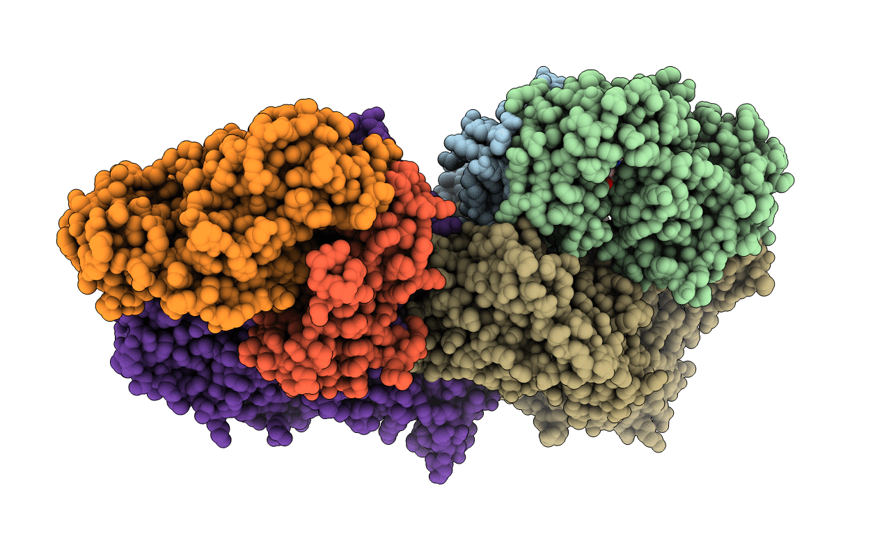
Structure of Xanthine Oxidase with 2-hydroxy-6-methylpurine
Biological Source:
Source Organism:
Bos taurus (Taxon ID: 9913)
Method Details:
Experimental Method:
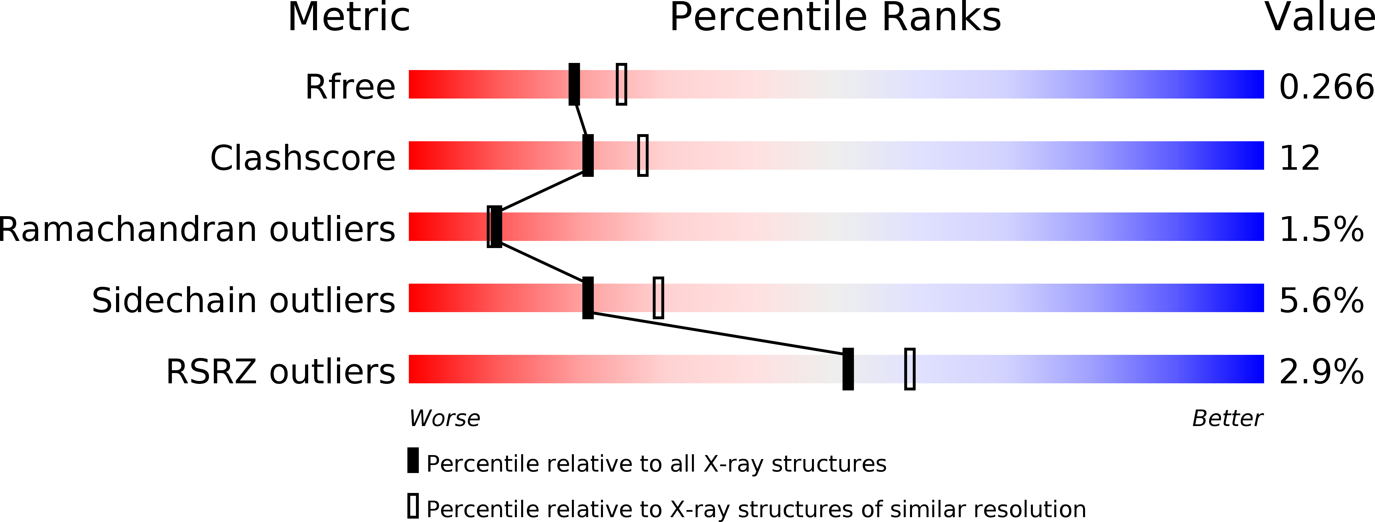
Resolution:
2.30 Å
R-Value Free:
0.26
R-Value Work:
0.19
R-Value Observed:
0.19
Space Group:
P 1 21 1
Macromolecular Entities
Polymer Type:polypeptide(L)
Molecule:xanthine oxidase
Gene (Uniprot):XDH
Chain IDs:A, D (auth: I)
Chain Length:219
Number of Molecules:2
Biological Source:Bos taurus
Polymer Type:polypeptide(L)
Molecule:xanthine oxidase
Gene (Uniprot):XDH
Chain IDs:B, E (auth: J)
Chain Length:350
Number of Molecules:2
Biological Source:Bos taurus
Polymer Type:polypeptide(L)
Molecule:xanthine oxidase
Gene (Uniprot):XDH
Chain IDs:C, F (auth: K)
Chain Length:763
Number of Molecules:2
Biological Source:Bos taurus
Primary Citation
Substrate orientation in xanthine oxidase: crystal structure of enzyme in reaction with 2-hydroxy-6-methylpurine.
J.Biol.Chem. 283 4818 4824 (2008)
PMID: 18063585 DOI: 10.1074/jbc.M707918200

Abstact

Xanthine oxidoreductase catalyzes the final two steps of purine catabolism and is involved in a variety of pathological states ranging from hyperuricemia to ischemia-reperfusion injury. The human enzyme is expressed primarily in its dehydrogenase form utilizing NAD+ as the final electron acceptor from the enzyme's flavin site but can exist as an oxidase that utilizes O2 for this purpose. Central to an understanding of the enzyme's function is knowledge of purine substrate orientation in the enzyme's molybdenum-containing active site. We report here the crystal structure of xanthine oxidase, trapped at the stage of a critical intermediate in the course of reaction with the slow substrate 2-hydroxy-6-methylpurine at 2.3A. This is the first crystal structure of a reaction intermediate with a purine substrate that is hydroxylated at its C8 position as is xanthine and confirms the structure predicted to occur in the course of the presently favored reaction mechanism. The structure also corroborates recent work suggesting that 2-hydroxy-6-methylpurine orients in the active site with its C2 carbonyl group interacting with Arg-880 and extends our hypothesis that xanthine binds opposite this orientation, with its C6 carbonyl positioned to interact with Arg-880 in stabilizing the MoV transition state.

Legend

Protein

Chemical

Disease

Primary Citation of related structures