3B99 image
Deposition Date 2007-11-03
Release Date 2007-11-20
Last Version Date 2023-11-01
Entry Detail
PDB ID:
3B99
Keywords:
Title:
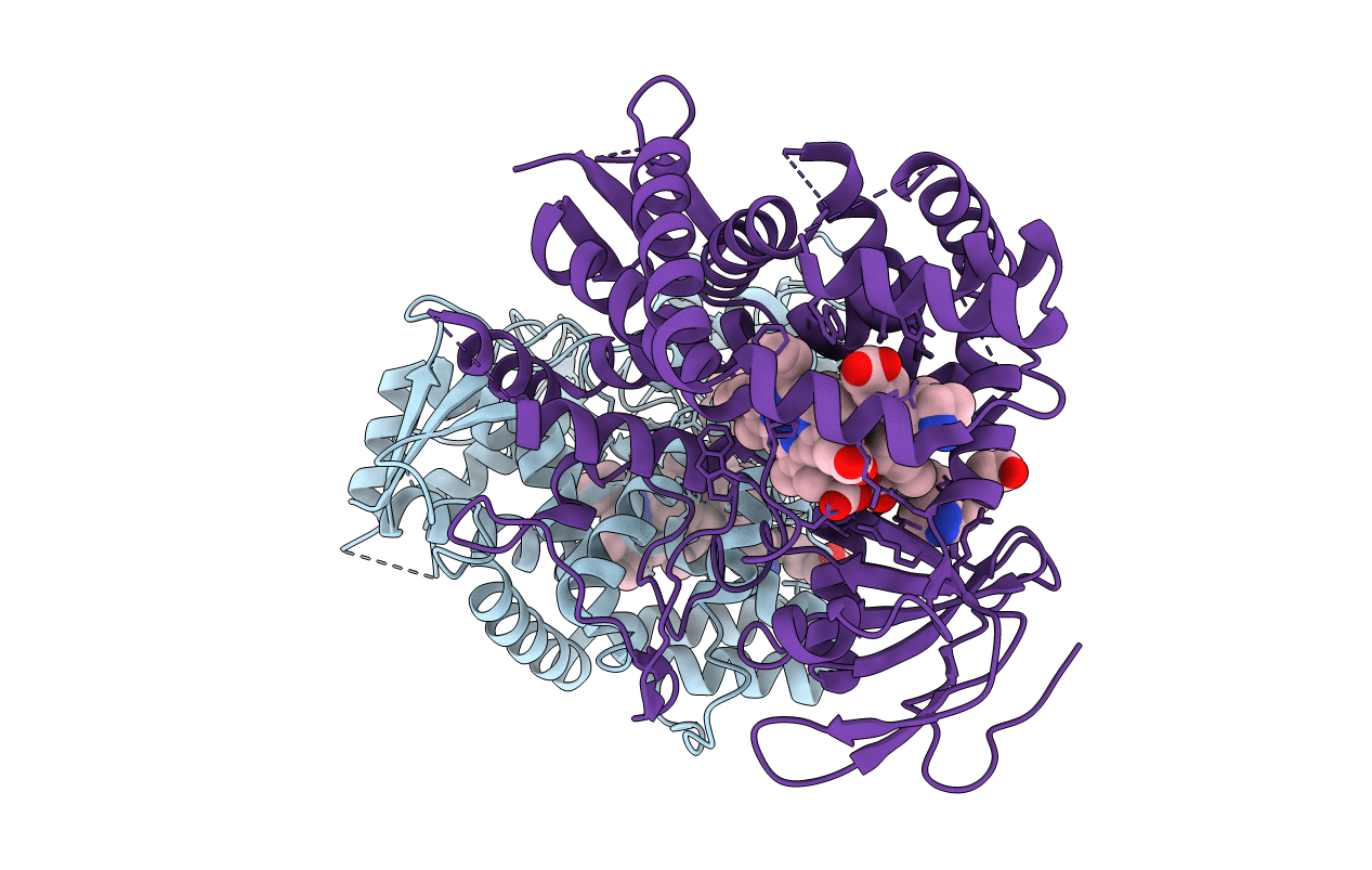
Crystal structure of zebrafish prostacyclin synthase (cytochrome P450 8A1) in complex with substrate analog U51605
Biological Source:
Source Organism:
Danio rerio (Taxon ID: 7955)
Method Details:
Experimental Method:
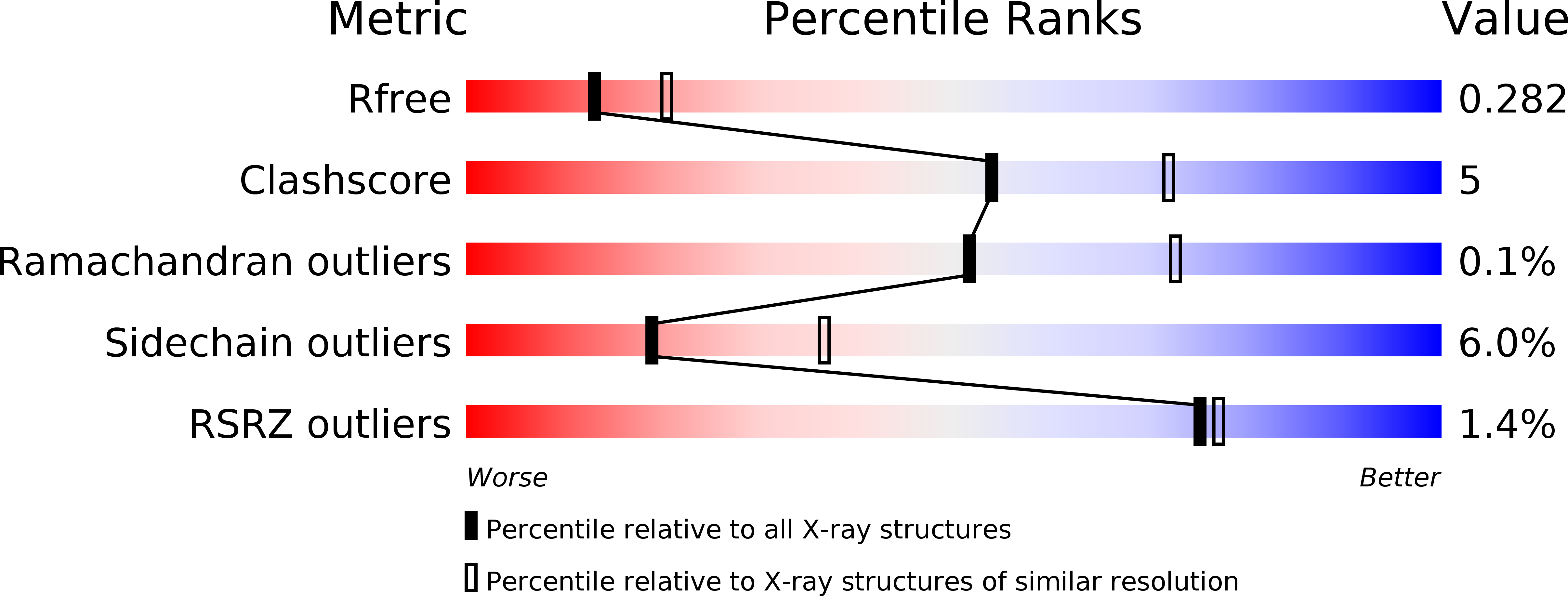
Resolution:
2.50 Å
R-Value Free:
0.29
R-Value Work:
0.21
R-Value Observed:
0.21
Space Group:
P 21 21 21
Macromolecular Entities
Structures with similar UniProt ID
Protein Blast
Polymer Type:polypeptide(L)
Molecule:Prostaglandin I2 synthase
Chain IDs:A, B
Chain Length:475
Number of Molecules:2
Biological Source:Danio rerio
Primary Citation
Structures of Prostacyclin Synthase and Its Complexes with Substrate Analog and Inhibitor Reveal a Ligand-specific Heme Conformation Change
J.Biol.Chem. 283 2917 2926 (2008)
PMID: 18032380 DOI: 10.1074/jbc.M707470200

Abstact

Prostacyclin synthase (PGIS) is a cytochrome P450 (P450) enzyme that catalyzes production of prostacyclin from prostaglandin H(2). PGIS is unusual in that it catalyzes an isomerization rather than a monooxygenation, which is typical of P450 enzymes. To understand the structural basis for prostacyclin biosynthesis in greater detail, we have determined the crystal structures of ligand-free, inhibitor (minoxidil)-bound and substrate analog U51605-bound PGIS. These structures demonstrate a stereo-specific substrate binding and suggest features of the enzyme that facilitate isomerization. Unlike most microsomal P450s, where large substrate-induced conformational changes take place at the distal side of the heme, conformational changes in PGIS are observed at the proximal side and in the heme itself. The conserved and extensive heme propionate-protein interactions seen in all other P450s, which are largely absent in the ligand-free PGIS, are recovered upon U51605 binding accompanied by water exclusion from the active site. In contrast, when minoxidil binds, the propionate-protein interactions are not recovered and water molecules are largely retained. These findings suggest that PGIS represents a divergent evolution of the P450 family, in which a heme barrier has evolved to ensure strict binding specificity for prostaglandin H(2), leading to a radical-mediated isomerization with high product fidelity. The U51605-bound structure also provides a view of the substrate entrance and product exit channels.

Legend

Protein

Chemical

Disease

Primary Citation of related structures