3B8M image
Deposition Date 2007-11-01
Release Date 2008-01-22
Last Version Date 2024-02-21
Entry Detail
PDB ID:
3B8M
Title:
Structure of FepE- Bacterial Polysaccharide Co-polymerase
Biological Source:
Source Organism:
Escherichia coli (Taxon ID: 562)
Method Details:
Experimental Method:
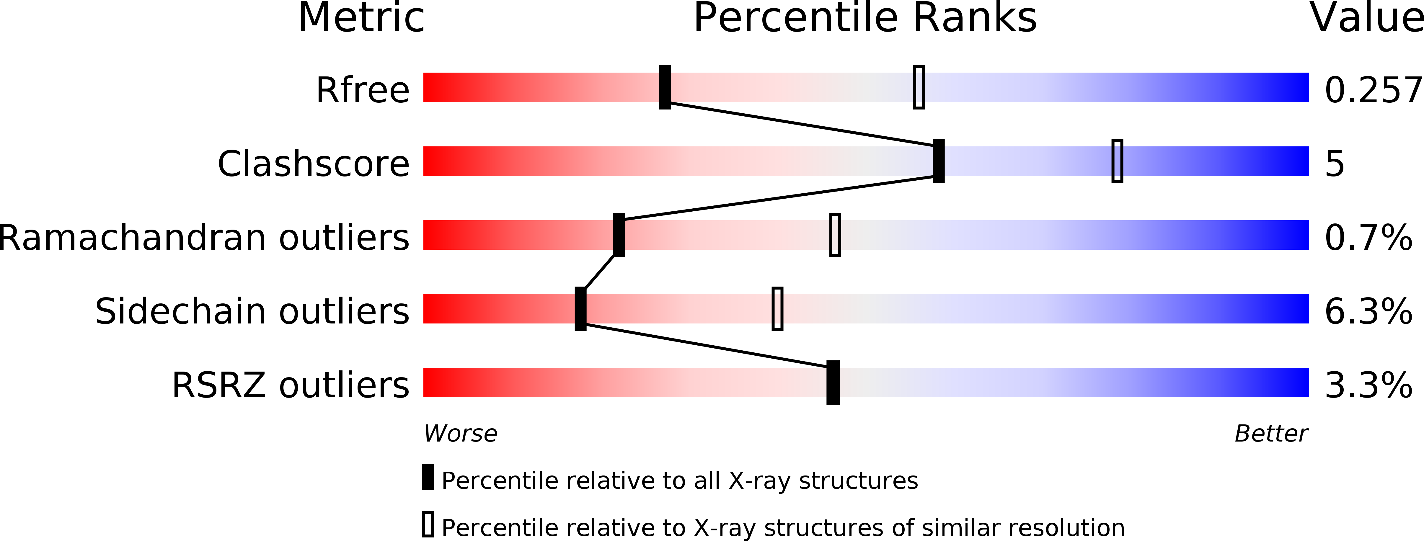
Resolution:
2.70 Å
R-Value Free:
0.26
R-Value Work:
0.22
R-Value Observed:
0.22
Space Group:
P 63 2 2
Macromolecular Entities
Polymer Type:polypeptide(L)
Molecule:Ferric enterobactin (Enterochelin) transport
Gene (Uniprot):fepE
Chain IDs:A, B, C
Chain Length:280
Number of Molecules:3
Biological Source:Escherichia coli
Primary Citation
Bacterial polysaccharide co-polymerases share a common framework for control of polymer length
Nat.Struct.Mol.Biol. 15 130 138 (2008)
PMID: 18204465 DOI: 10.1038/nsmb.1374

Abstact

The chain length distribution of complex polysaccharides present on the bacterial surface is determined by polysaccharide co-polymerases (PCPs) anchored in the inner membrane. We report crystal structures of the periplasmic domains of three PCPs that impart substantially different chain length distributions to surface polysaccharides. Despite very low sequence similarities, they have a common protomer structure with a long central alpha-helix extending 100 A into the periplasm. The protomers self-assemble into bell-shaped oligomers of variable sizes, with a large internal cavity. Electron microscopy shows that one of the full-length PCPs has a similar organization as that observed in the crystal for its periplasmic domain alone. Functional studies suggest that the top of the PCP oligomers is an important region for determining polysaccharide modal length. These structures provide a detailed view of components of the bacterial polysaccharide assembly machinery.

Legend

Protein

Chemical

Disease

Primary Citation of related structures