3B7E image
Entry Detail
PDB ID:
3B7E
Keywords:
Title:
Neuraminidase of A/Brevig Mission/1/1918 H1N1 strain in complex with zanamivir
Biological Source:
Source Organism:
Host Organism:
PDB Version:
Deposition Date:
2007-10-30
Release Date:
2008-10-07
Method Details:
Experimental Method:
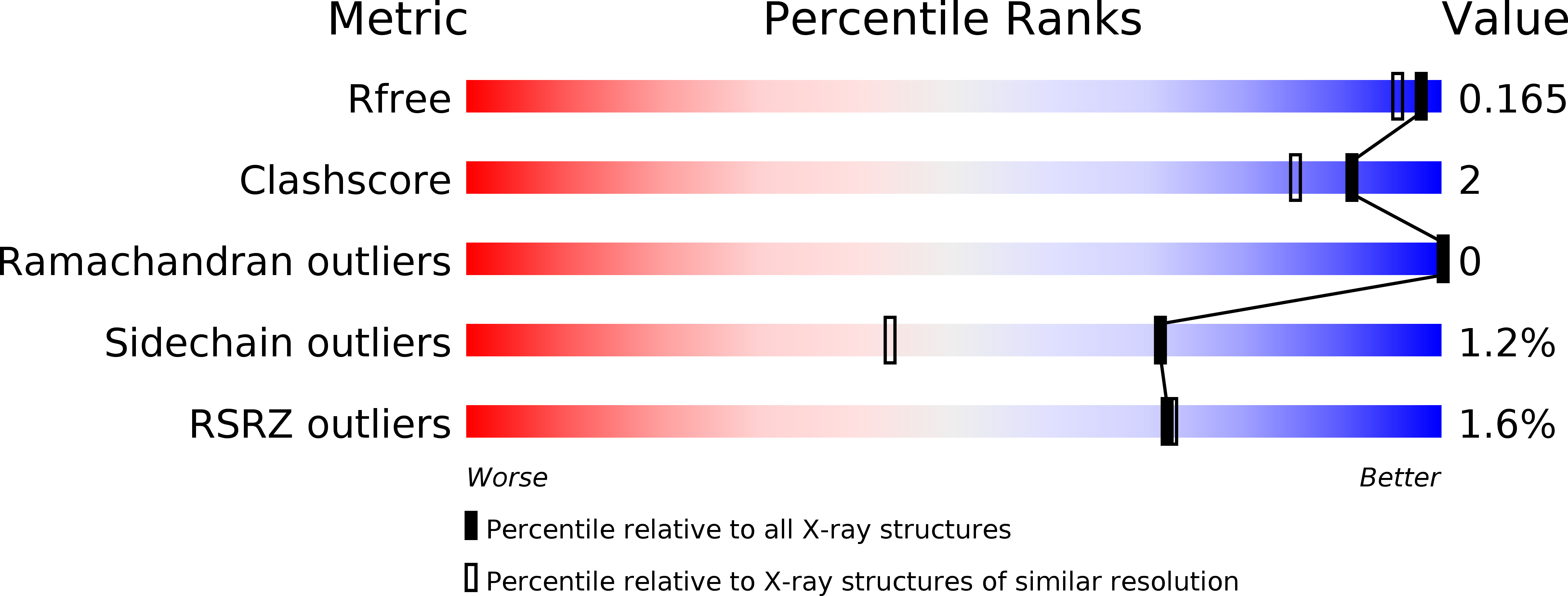
Resolution:
1.45 Å
R-Value Free:
0.16
R-Value Work:
0.14
R-Value Observed:
0.14
Space Group:
C 2 2 21
Macromolecular Entities
Polymer Type:polypeptide(L)
Description:Neuraminidase
Chain IDs:A, B
Chain Length:385
Number of Molecules:2
Biological Source:Influenza A virus
Modified Residue
Compound ID Chain ID Parent Comp ID Details 2D Image
ASN A ASN GLYCOSYLATION SITE
Primary Citation
Structural characterization of the 1918 influenza virus H1N1 neuraminidase
J.Virol. 82 10493 10501 (2008)
PMID: 18715929 DOI: 10.1128/JVI.00959-08

Abstact

Influenza virus neuraminidase (NA) plays a crucial role in facilitating the spread of newly synthesized virus in the host and is an important target for controlling disease progression. The NA crystal structure from the 1918 "Spanish flu" (A/Brevig Mission/1/18 H1N1) and that of its complex with zanamivir (Relenza) at 1.65-A and 1.45-A resolutions, respectively, corroborated the successful expression of correctly folded NA tetramers in a baculovirus expression system. An additional cavity adjacent to the substrate-binding site is observed in N1, compared to N2 and N9 NAs, including H5N1. This cavity arises from an open conformation of the 150 loop (Gly147 to Asp151) and appears to be conserved among group 1 NAs (N1, N4, N5, and N8). It closes upon zanamivir binding. Three calcium sites were identified, including a novel site that may be conserved in N1 and N4. Thus, these high-resolution structures, combined with our recombinant expression system, provide new opportunities to augment the limited arsenal of therapeutics against influenza.

Legend

Protein

Chemical

Disease

Primary Citation of related structures