Deposition Date
2007-10-25
Release Date
2007-12-04
Last Version Date
2024-02-21
Entry Detail
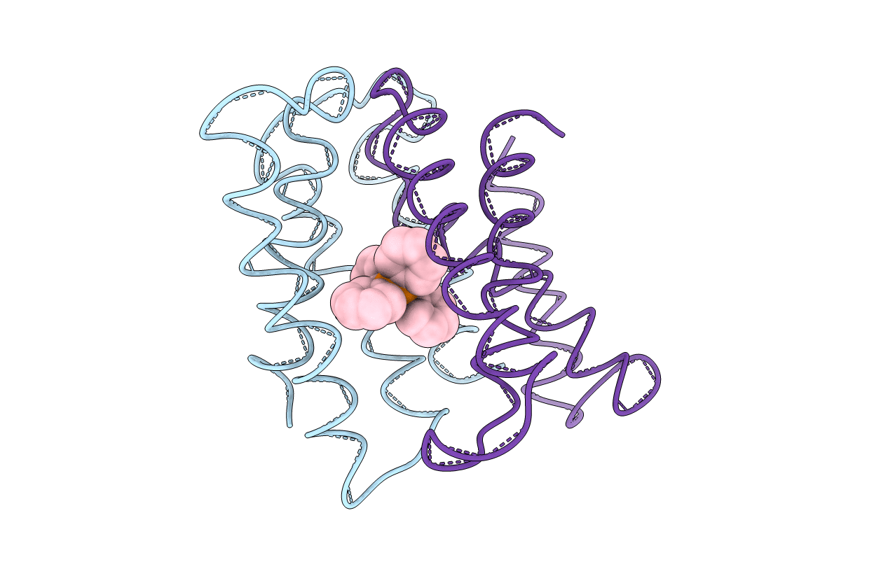
PDB ID:
3B5D
Keywords:
Title:
EmrE multidrug transporter in complex with TPP, C2 crystal form
Biological Source:
Source Organism(s):
Escherichia coli K12 (Taxon ID: 83333)
Expression System(s):
Method Details:
Experimental Method:
Resolution:
3.80 Å
R-Value Free:
0.36
R-Value Work:
0.32
Space Group:
C 1 2 1