3B2R image
Deposition Date 2007-10-19
Release Date 2008-05-20
Last Version Date 2024-04-03
Entry Detail
PDB ID:
3B2R
Keywords:
Title:
Crystal Structure of PDE5A1 catalytic domain in complex with Vardenafil
Biological Source:
Source Organism:
Homo sapiens (Taxon ID: )
Host Organism:
Method Details:
Experimental Method:
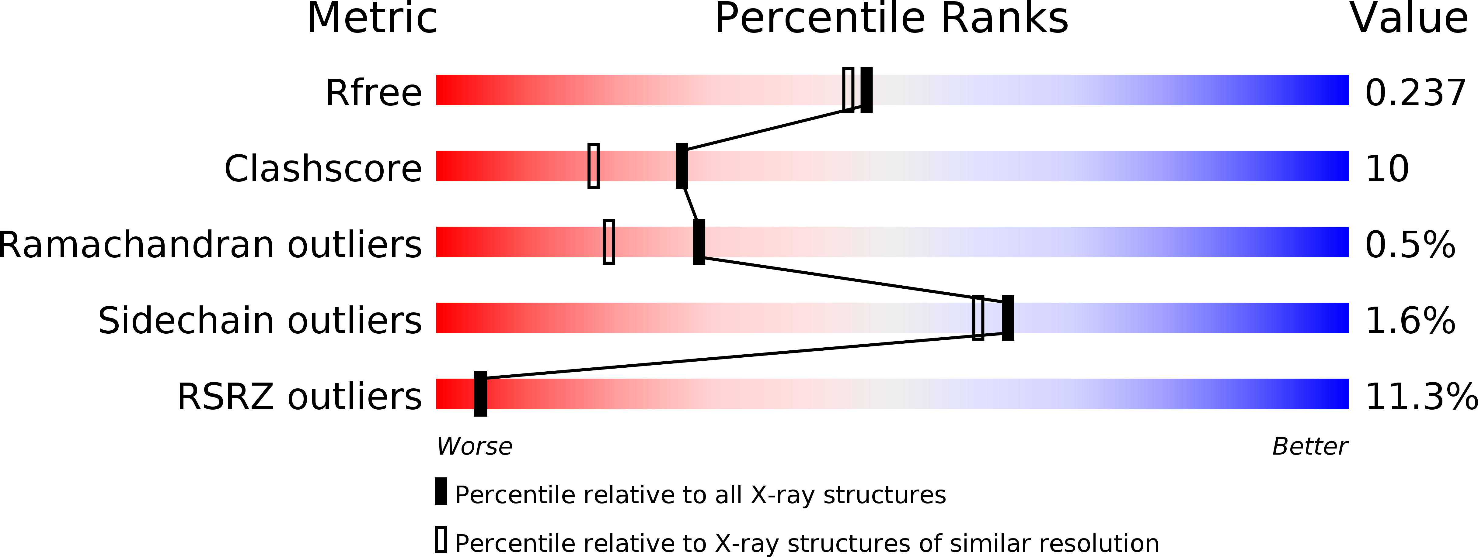
Resolution:
2.07 Å
R-Value Free:
0.24
R-Value Work:
0.21
Space Group:
P 21 21 21
Macromolecular Entities
Polymer Type:polypeptide(L)
Molecule:cGMP-specific 3',5'-cyclic phosphodiesterase
Gene (Uniprot):PDE5A
Chain IDs:A, B
Chain Length:330
Number of Molecules:2
Biological Source:Homo sapiens
Ligand Molecules
Primary Citation
Conformational variations of both phosphodiesterase-5 and inhibitors provide the structural basis for the physiological effects of vardenafil and sildenafil.
Mol.Pharmacol. 73 104 110 (2008)
PMID: 17959709 DOI: 10.1124/mol.107.040212

Abstact

Vardenafil has higher affinity to phosphodiesterase-5 (PDE5) than sildenafil and lower administered dosage for the treatment of erectile dysfunction. However, the molecular basis for these differences is puzzling because two drugs have similar chemical structures. Reported here is a crystal structure of the fully active and nonmutated PDE5A1 catalytic domain in complex with vardenafil. The structure shows that the conformation of the H-loop in the PDE5A1-vardenafil complex is different from those of any known structures of the unliganded PDE5 and its complexes with the inhibitors. In addition, the molecular configuration of vardenafil differs from that of sildenafil when bound to PDE5. It is noteworthy that the binding of vardenafil causes loss of the divalent metal ions that have been observed in all the previously published PDE structures. The conformational variation of both PDE5 and the inhibitors provides structural insight into the different potencies of the drugs.

Legend

Protein

Chemical

Disease

Primary Citation of related structures