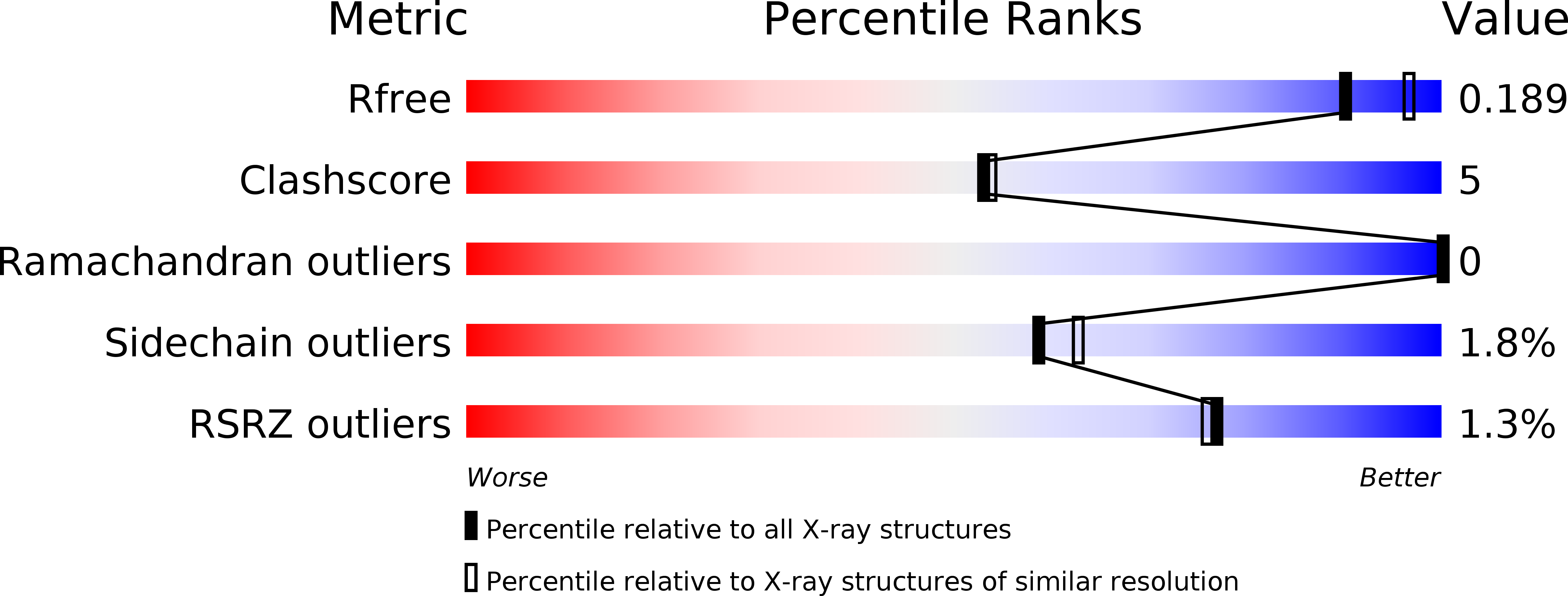
3B2P image
Deposition Date 2007-10-18
Release Date 2008-05-06
Last Version Date 2023-08-30
Entry Detail
PDB ID:
3B2P
Keywords:
Title:
Crystal structure of E. coli Aminopeptidase N in complex with arginine
Biological Source:
Source Organism:
Method Details:
Experimental Method:
Resolution:
2.00 Å
R-Value Free:
0.18
R-Value Work:
0.15
R-Value Observed:
0.15
Space Group:
P 31 2 1
Macromolecular Entities
Polymer Type:polypeptide(L)
Molecule:Aminopeptidase N
Gene (Uniprot):pepN
Chain IDs:A
Chain Length:891
Number of Molecules:1
Biological Source:Escherichia coli K12
Primary Citation
Structural basis for the unusual specificity of Escherichia coli aminopeptidase N.
Biochemistry 47 5303 5311 (2008)
PMID: 18416562 DOI: 10.1021/bi7022333

Abstact

Aminopeptidase N from Escherichia coli is a M1 class aminopeptidase with the active-site region related to that of thermolysin. The enzyme has unusual specificity, cleaving adjacent to the large, nonpolar amino acids Phe and Tyr but also cleaving next to the polar residues Lys and Arg. To try to understand the structural basis for this pattern of hydrolysis, the structure of the enzyme was determined in complex with the amino acids L-arginine, L-lysine, L-phenylalanine, L-tryptophan, and L-tyrosine. These amino acids all bind with their backbone atoms close to the active-site zinc ion and their side chain occupying the S1 subsite. This subsite is in the form of a cylinder, about 10 A in cross-section and 12 A in length. The bottom of the cylinder includes the zinc ion and a number of polar side chains that make multiple hydrogen-bonding and other interactions with the alpha-amino group and the alpha-carboxylate of the bound amino acid. The walls of the S1 cylinder are hydrophobic and accommodate the nonpolar or largely nonpolar side chains of Phe and Tyr. The top of the cylinder is polar in character and includes bound water molecules. The epsilon-amino group of the bound lysine side chain and the guanidinium group of arginine both make multiple hydrogen bonds to this part of the S1 site. At the same time, the hydrocarbon part of the lysine and arginine side chains is accommodated within the nonpolar walls of the S1 cylinder. This combination of hydrophobic and hydrophilic binding surfaces explains the ability of ePepN to cleave Lys, Arg, Phe, and Tyr. Another favored substrate has Ala at the P1 position. The short, nonpolar side chain of this residue can clearly be bound within the hydrophobic part of the S1 cylinder, but the reason for its facile hydrolysis remains uncertain.

Legend

Protein

Chemical

Disease

Primary Citation of related structures