Deposition Date
2011-07-02
Release Date
2011-10-26
Last Version Date
2024-10-23
Entry Detail
PDB ID:
3B1F
Keywords:
Title:
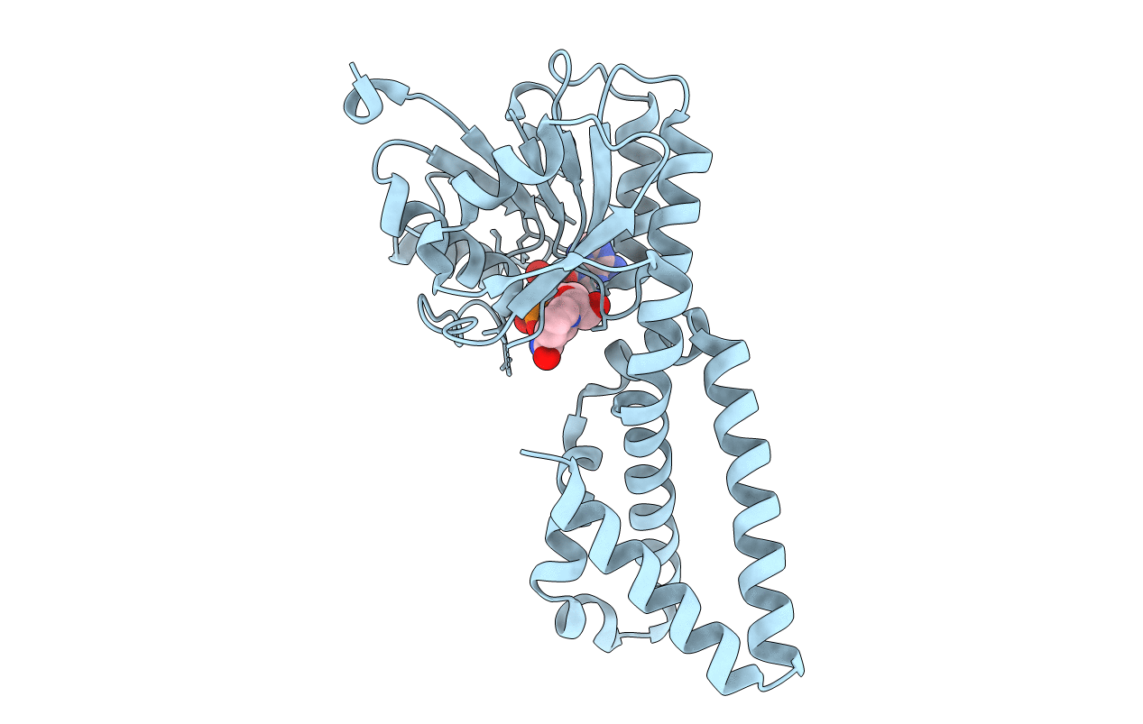
Crystal structure of prephenate dehydrogenase from Streptococcus mutans
Biological Source:
Source Organism(s):
Streptococcus mutans (Taxon ID: 1309)
Expression System(s):
Method Details:
Experimental Method:
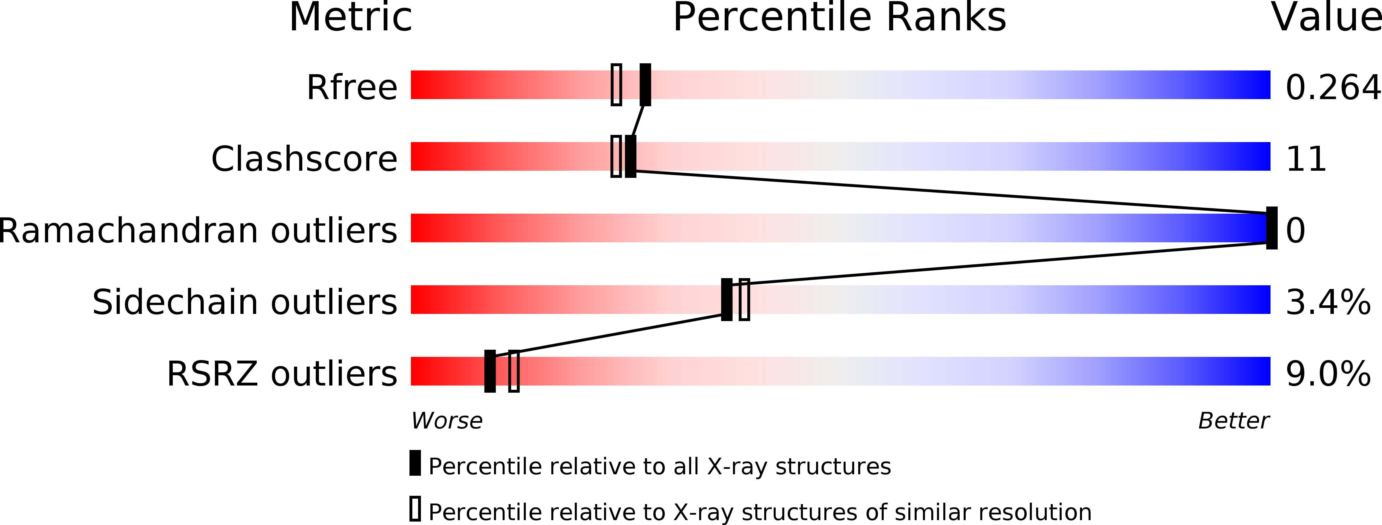
Resolution:
2.10 Å
R-Value Free:
0.23
R-Value Work:
0.19
R-Value Observed:
0.19
Space Group:
C 2 2 21