Deposition Date
2011-06-14
Release Date
2011-12-14
Last Version Date
2024-10-23
Entry Detail
PDB ID:
3B0V
Keywords:
Title:
tRNA-dihydrouridine synthase from Thermus thermophilus in complex with tRNA
Biological Source:
Source Organism(s):
Thermus thermophilus (Taxon ID: 300852)
Expression System(s):
Method Details:
Experimental Method:
Resolution:
3.51 Å
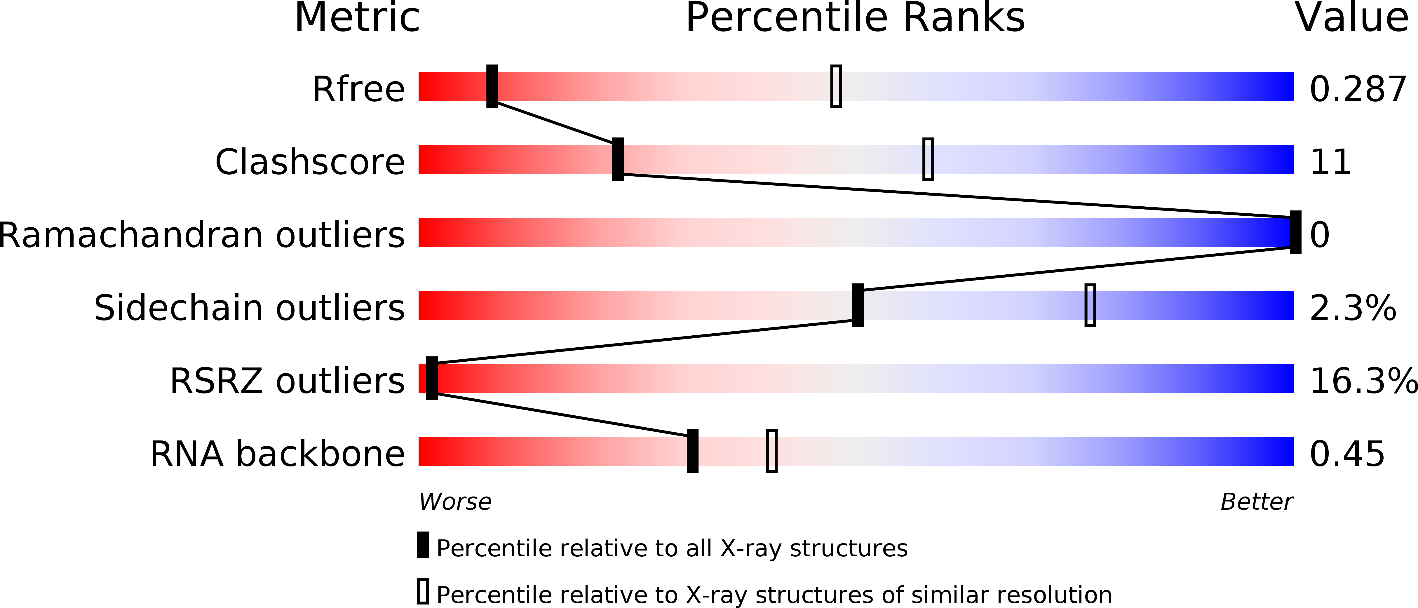
R-Value Free:
0.32
R-Value Work:
0.30
Space Group:
P 41 21 2