3B09 image
Deposition Date 2011-06-07
Release Date 2012-04-18
Last Version Date 2024-10-30
Entry Detail
PDB ID:
3B09
Keywords:
Title:
Crystal structure of the N-domain of FKBP22 from Shewanella sp. SIB1
Biological Source:
Source Organism:
Shewanella (Taxon ID: 117911)
Host Organism:
Method Details:
Experimental Method:
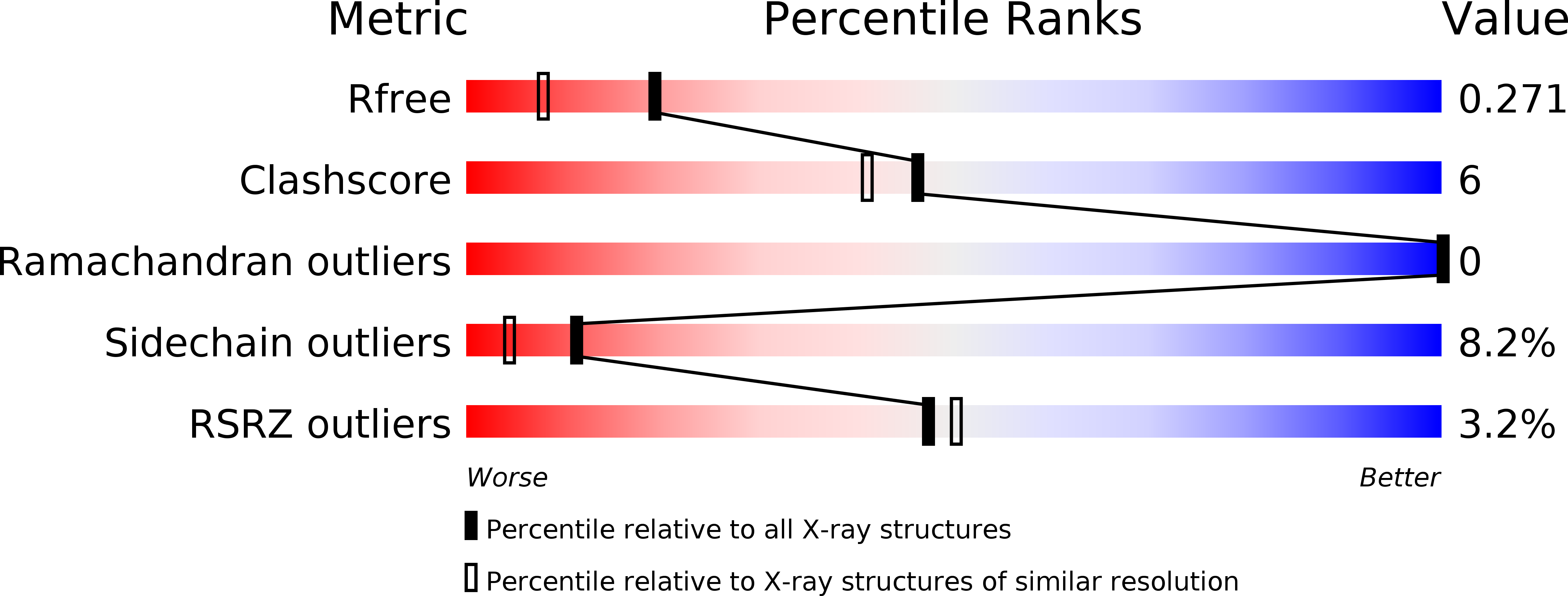
Resolution:
1.90 Å
R-Value Free:
0.27
R-Value Work:
0.23
R-Value Observed:
0.23
Space Group:
P 32 2 1
Macromolecular Entities
Structures with similar UniProt ID
Protein Blast
Polymer Type:polypeptide(L)
Molecule:Peptidyl-prolyl cis-trans isomerase
Gene (Uniprot):fklB
Chain IDs:A
Chain Length:88
Number of Molecules:1
Biological Source:Shewanella
Modified Residue
Compound ID Chain ID Parent Comp ID Details 2D Image
MSE A MET SELENOMETHIONINE
Primary Citation
Crystal structure of N-domain of FKBP22 from Shewanella sp. SIB1: dimer dissociation by disruption of Val-Leu knot
Protein Sci. 20 1755 1764 (2011)
PMID: 21837652 DOI: 10.1002/pro.714

Abstact

FK506-binding protein 22 (FKBP22) from the psychrotophic bacterium Shewanella sp. SIB1 (SIB1 FKBP22) is a homodimeric protein with peptidyl prolyl cis-trans isomerase (PPIase) activity. Each monomer consists of the N-terminal domain responsible for dimerization and C-terminal catalytic domain. To reveal interactions at the dimer interface of SIB1 FKBP22, the crystal structure of the N-domain of SIB1 FKBP22 (SN-FKBP22, residues 1-68) was determined at 1.9 Å resolution. SN-FKBP22 forms a dimer, in which each monomer consists of three helices (α1, α2, and α3N). In the dimer, two monomers have head-to-head interactions, in which residues 8-64 of one monomer form tight interface with the corresponding residues of the other. The interface is featured by the presence of a Val-Leu knot, in which Val37 and Leu41 of one monomer interact with Val41 and Leu37 of the other, respectively. To examine whether SIB1 FKBP22 is dissociated into the monomers by disruption of this knot, the mutant protein V37R/L41R-FKBP22, in which Val37 and Leu41 of SIB1 FKBP22 are simultaneously replaced by Arg, was constructed and biochemically characterized. This mutant protein was indistinguishable from the SIB1 FKBP22 derivative lacking the N-domain in oligomeric state, far-UV CD spectrum, thermal denaturation curve, PPIase activity, and binding ability to a folding intermediate of protein, suggesting that the N-domain of V37R/L41R-FKBP22 is disordered. We propose that a Val-Leu knot at the dimer interface of SIB1 FKBP22 is important for dimerization and dimerization is required for folding of the N-domain.

Legend

Protein

Chemical

Disease

Primary Citation of related structures
Feedback Form
Name
Email
Institute
Feedback