3AZD image
Deposition Date 2011-05-23
Release Date 2011-10-19
Last Version Date 2023-11-01
Entry Detail
PDB ID:
3AZD
Title:
Crystal structure of tropomyosin N-terminal fragment at 0.98A resolution
Biological Source:
Source Organism:
Method Details:
Experimental Method:
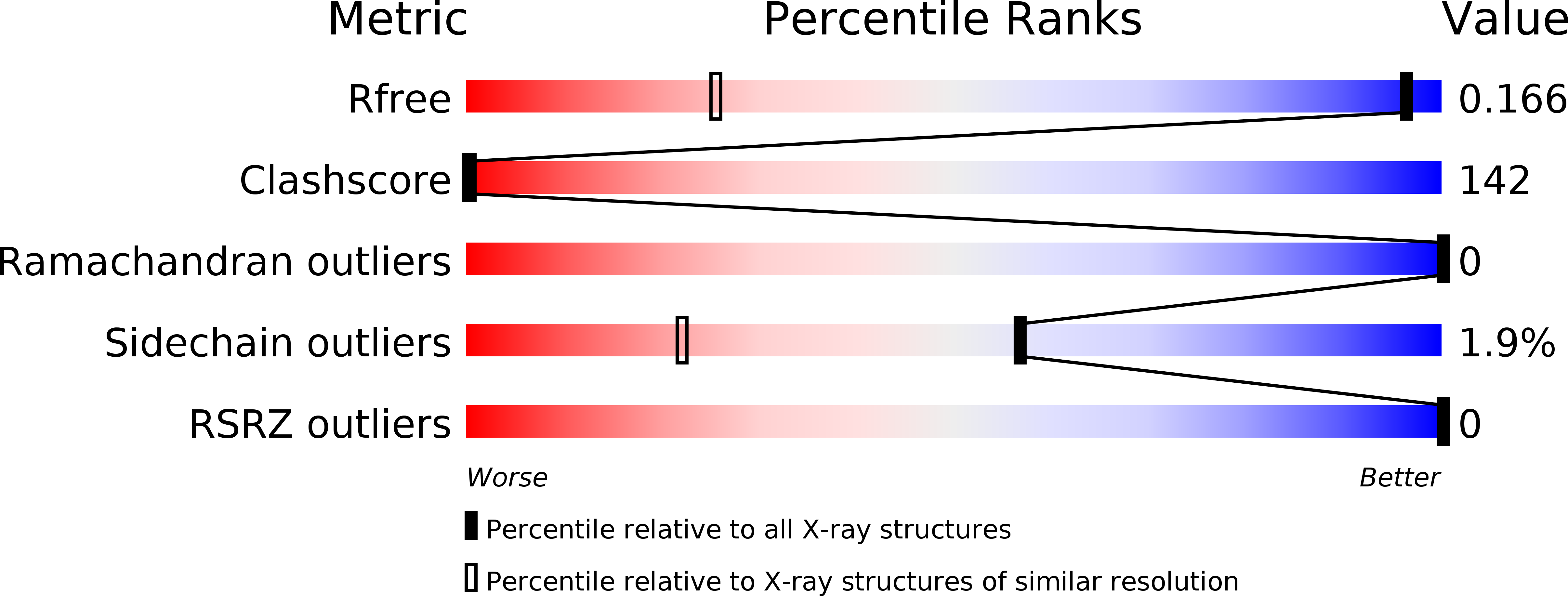
Resolution:
0.98 Å
R-Value Free:
0.17
R-Value Work:
0.16
R-Value Observed:
0.16
Space Group:
P 31
Macromolecular Entities
Polymer Type:polypeptide(L)
Molecule:short alpha-tropomyosin,transcription factor GCN4
Gene (Uniprot):GCN4
Chain IDs:A, B
Chain Length:37
Number of Molecules:2
Biological Source:Rattus norvegicus, Saccharomyces cerevisiae S288c
Primary Citation
Structure of a tropomyosin N-terminal fragment at 0.98 A resolution
Acta Crystallogr.,Sect.D 67 822 825 (2011)
PMID: 21904035 DOI: 10.1107/S090744491102645X

Abstact

Tropomyosin (TM) is an elongated two-chain protein that binds along actin filaments. Important binding sites are localized in the N-terminus of tropomyosin. The structure of the N-terminus of the long muscle α-TM has been solved by both NMR and X-ray crystallography. Only the NMR structure of the N-terminus of the short nonmuscle α-TM is available. Here, the crystal structure of the N-terminus of the short nonmuscle α-TM (αTm1bZip) at a resolution of 0.98 Å is reported, which was solved from crystals belonging to space group P3(1) with unit-cell parameters a = b = 33.00, c = 52.03 Å, α = β = 90, γ = 120°. The first five N-terminal residues are flexible and residues 6-35 form an α-helical coiled coil. The overall fold and the secondary structure of the crystal structure of αTM1bZip are highly similar to the NMR structure and the atomic coordinates of the corresponding C(α) atoms between the two structures superimpose with a root-mean-square deviation of 0.60 Å. The crystal structure validates the NMR structure, with the positions of the side chains being determined precisely in our structure.

Legend

Protein

Chemical

Disease

Primary Citation of related structures