3AYU image
Deposition Date 2011-05-17
Release Date 2011-08-03
Last Version Date 2024-03-13
Entry Detail
PDB ID:
3AYU
Title:
Crystal structure of MMP-2 active site mutant in complex with APP-drived decapeptide inhibitor
Biological Source:
Source Organism:
Homo sapiens (Taxon ID: 9606)
Host Organism:
Method Details:
Experimental Method:
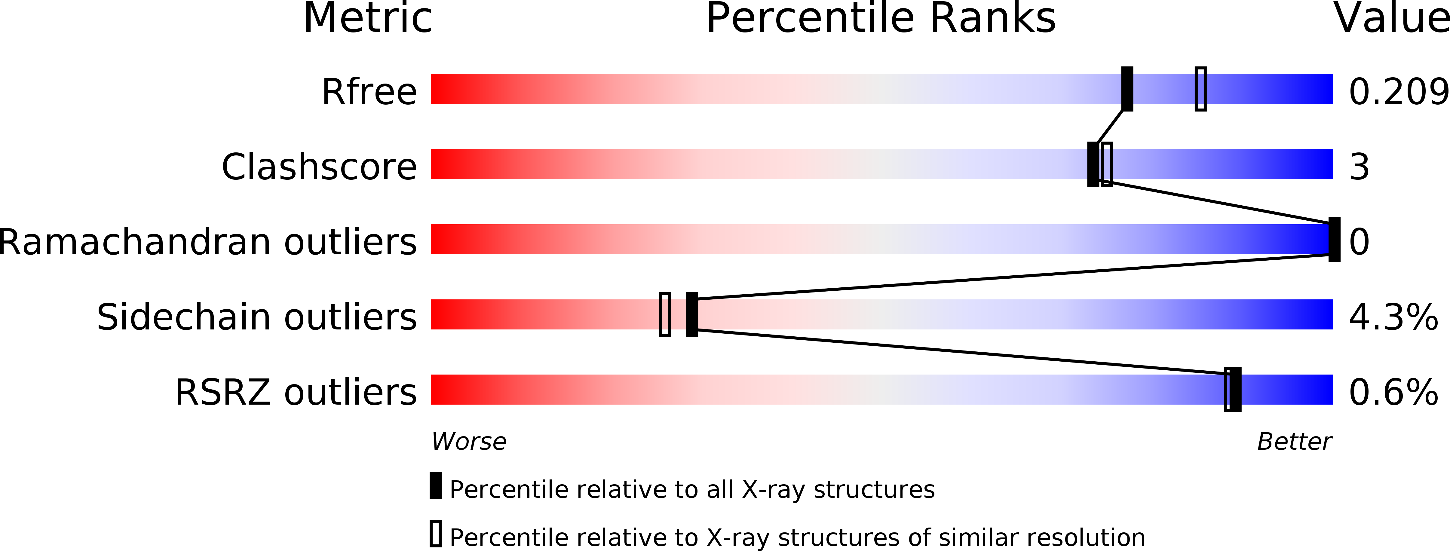
Resolution:
2.00 Å
R-Value Free:
0.20
R-Value Work:
0.16
R-Value Observed:
0.16
Space Group:
P 21 21 21
Macromolecular Entities
Structures with similar UniProt ID
Protein Blast
Polymer Type:polypeptide(L)
Molecule:72 kDa type IV collagenase
Gene (Uniprot):MMP2
Mutagens:E108K, Q110V, E121A
Chain IDs:A
Chain Length:167
Number of Molecules:1
Biological Source:Homo sapiens
Structures with similar UniProt ID
Protein Blast
Polymer Type:polypeptide(L)
Molecule:Amyloid beta A4 protein
Gene (Uniprot):APP
Chain IDs:B
Chain Length:10
Number of Molecules:1
Biological Source:Homo sapiens
Primary Citation
Structural basis for matrix metalloproteinase-2 (MMP-2)-selective inhibitory action of {beta}-amyloid precursor protein-derived inhibitor
J.Biol.Chem. ? ? ? (2011)
PMID: 21813640 DOI: 10.1074/jbc.M111.264176

Abstact

Unlike other synthetic or physiological inhibitors for matrix metalloproteinases (MMPs), the β-amyloid precursor protein-derived inhibitory peptide (APP-IP) having an ISYGNDALMP sequence has a high selectivity toward MMP-2. Our previous study identified amino acid residues of MMP-2 essential for its selective inhibition by APP-IP and demonstrated that the N to C direction of the decapeptide inhibitor relative to the substrate-binding cleft of MMP-2 is opposite that of substrate. However, detailed interactions between the two molecules remained to be clarified. Here, we determined the crystal structure of the catalytic domain of MMP-2 in complex with APP-IP. We found that APP-IP in the complex is indeed embedded into the substrate-binding cleft of the catalytic domain in the N to C direction opposite that of substrate. With the crystal structure, it was first clarified that the aromatic side chain of Tyr(3) of the inhibitor is accommodated into the S1' pocket of the protease, and the carboxylate group of Asp(6) of APP-IP coordinates bidentately to the catalytic zinc of the enzyme. The Ala(7) to Pro(10) and Tyr(3) to Ile(1) strands of the inhibitor extend into the nonprime and the prime sides of the cleft, respectively. Therefore, the decapeptide inhibitor has long range contact with the substrate-binding cleft of the protease. This mode of interaction is probably essential for the high MMP-2 selectivity of the inhibitor because MMPs share a common architecture in the vicinity of the catalytic center, but whole structures of their substrate-binding clefts have sufficient variety for the inhibitor to distinguish MMP-2 from other MMPs.

Legend

Protein

Chemical

Disease

Primary Citation of related structures