
Deposition Date
2011-03-19
Release Date
2011-05-04
Last Version Date
2024-10-16
Entry Detail
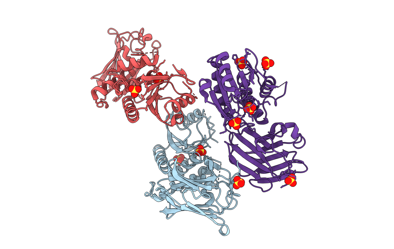
PDB ID:
3AWG
Keywords:
Title:
Crystal structure of Pten-like domain of Ci-VSP G356A mutant (248-576)
Biological Source:
Source Organism(s):
Ciona intestinalis (Taxon ID: 7719)
Expression System(s):
Method Details:
Experimental Method:
Resolution:
2.39 Å
R-Value Free:
0.25
R-Value Work:
0.19
R-Value Observed:
0.19
Space Group:
P 21 21 2
