3AV3 image
Entry Detail
PDB ID:
3AV3
Keywords:
Title:
Crystal structure of glycinamide ribonucleotide transformylase 1 from Geobacillus kaustophilus
Biological Source:
Source Organism:
Host Organism:
PDB Version:
Deposition Date:
2011-02-18
Release Date:
2012-03-07
Method Details:
Experimental Method:
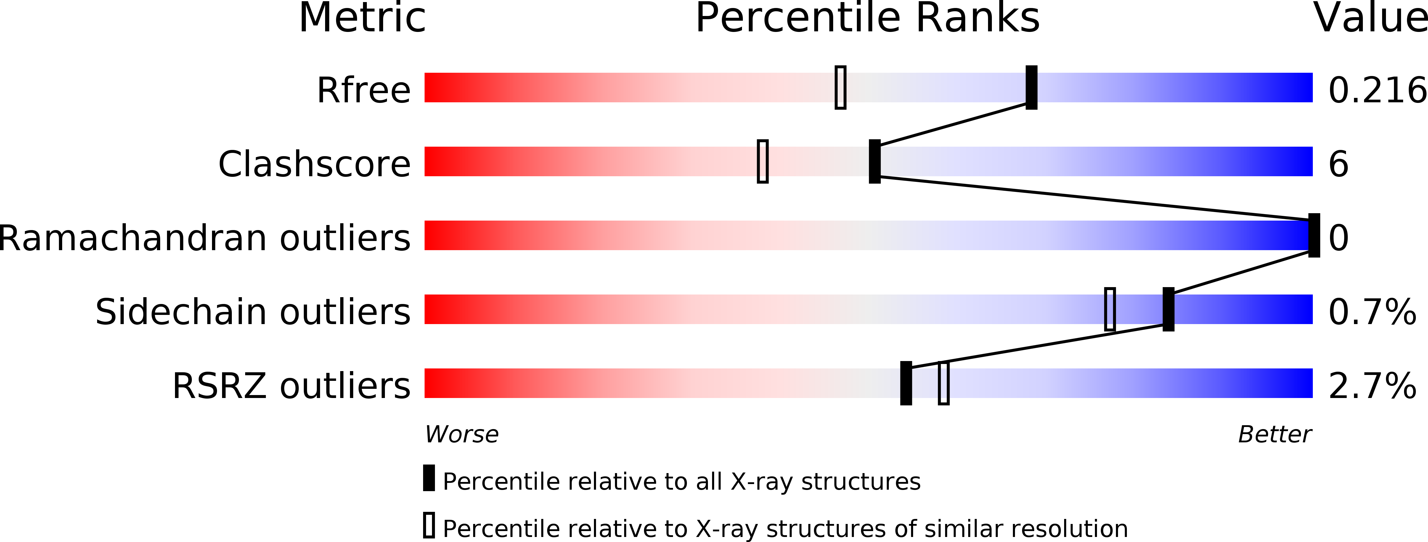
Resolution:
1.70 Å
R-Value Free:
0.21
R-Value Work:
0.19
R-Value Observed:
0.19
Space Group:
C 2 2 21
Macromolecular Entities
Polymer Type:polypeptide(L)
Description:Phosphoribosylglycinamide formyltransferase
Chain IDs:A
Chain Length:212
Number of Molecules:1
Biological Source:Geobacillus kaustophilus
Modified Residue
Compound ID Chain ID Parent Comp ID Details 2D Image
MSE A MET SELENOMETHIONINE
Ligand Molecules
Primary Citation
Structures and reaction mechanisms of the two related enzymes, PurN and PurU.
J.Biochem. 154 569 579 (2013)
PMID: 24108189 DOI: 10.1093/jb/mvt090

Abstact

The crystal structures of glycinamide ribonucleotide transformylases (PurNs) from Aquifex aeolicus (Aa), Geobacillus kaustophilus (Gk) and Symbiobacterium toebii (St), and of formyltetrahydrofolate hydrolase (PurU) from Thermus thermophilus (Tt) were determined. The monomer structures of the determined PurN and PurU were very similar to the known structure of PurN, but oligomeric states were different; AaPurN and StPurN formed dimers, GkPurN formed monomer and PurU formed tetramer in the crystals. PurU had a regulatory ACT domain in its N-terminal side. So far several structures of PurUs have been determined, yet, the mechanisms of the catalysis and the regulation of PurU have not been elucidated. We, therefore, modelled ligand-bound structures of PurN and PurU, and performed molecular dynamics simulations to elucidate the reaction mechanisms. The evolutionary relationship of the two enzymes is discussed based on the comparisons of the structures and the catalytic mechanisms.

Legend

Protein

Chemical

Disease

Primary Citation of related structures