3AUJ image
Deposition Date 2011-02-07
Release Date 2012-02-22
Last Version Date 2023-11-01
Entry Detail
PDB ID:
3AUJ
Keywords:
Title:
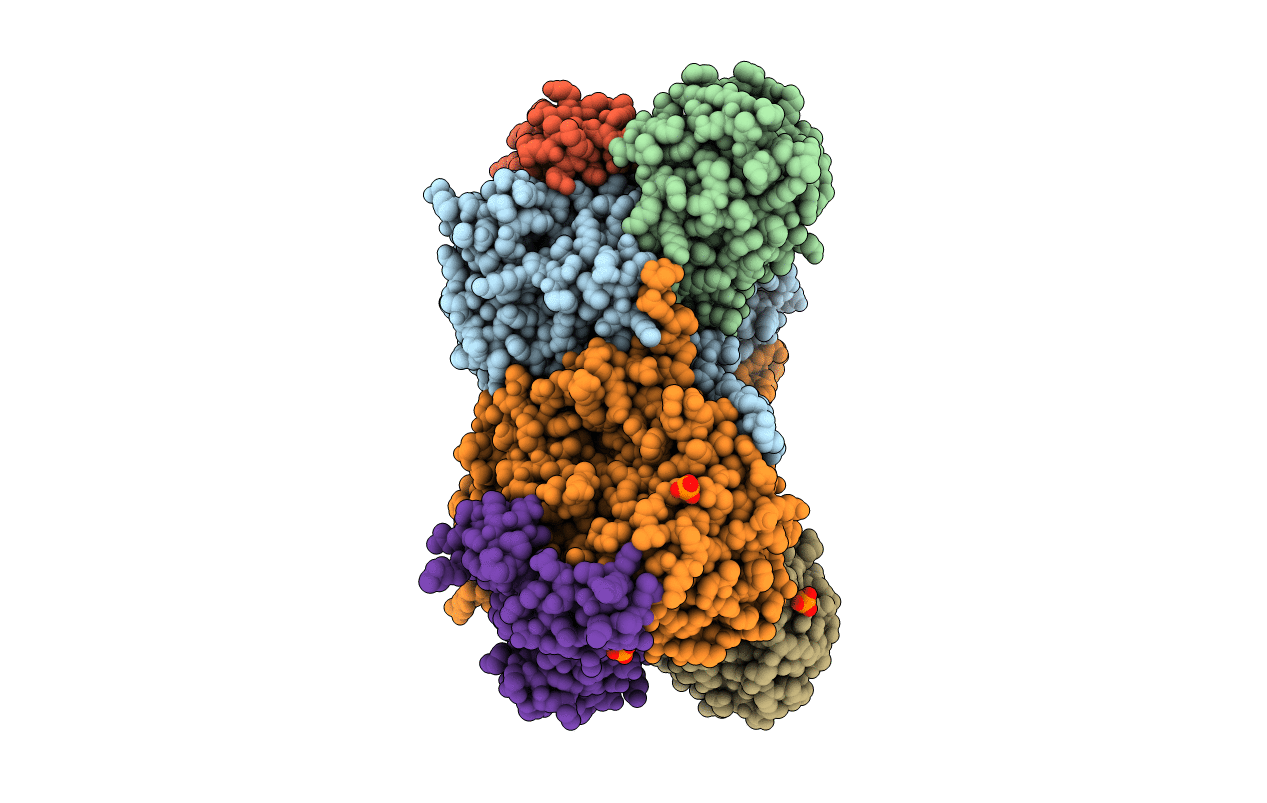
Structure of diol dehydratase complexed with glycerol
Biological Source:
Source Organism:
Host Organism:
Method Details:
Experimental Method:
Resolution:
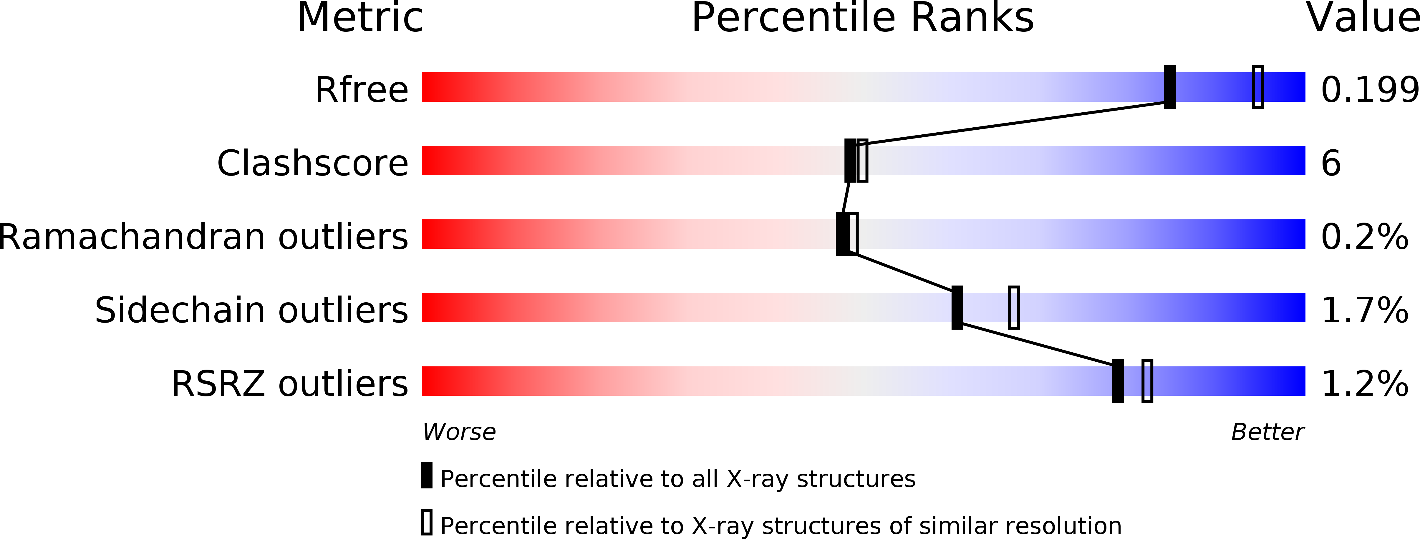
2.10 Å
R-Value Free:
0.19
R-Value Work:
0.18
R-Value Observed:
0.18
Space Group:
C 1 2 1
Macromolecular Entities
Polymer Type:polypeptide(L)
Molecule:Diol dehydrase alpha subunit
Gene (Uniprot):pddA
Chain IDs:A, D (auth: L)
Chain Length:554
Number of Molecules:2
Biological Source:Klebsiella oxytoca
Polymer Type:polypeptide(L)
Molecule:Diol dehydrase beta subunit
Gene (Uniprot):pddB
Chain IDs:B, E
Chain Length:224
Number of Molecules:2
Biological Source:Klebsiella oxytoca
Polymer Type:polypeptide(L)
Molecule:Diol dehydrase gamma subunit
Gene (Uniprot):pddC
Chain IDs:C (auth: G), F (auth: M)
Chain Length:173
Number of Molecules:2
Biological Source:Klebsiella oxytoca
Primary Citation
Redesign of coenzyme B(12) dependent diol dehydratase to be resistant to the mechanism-based inactivation by glycerol and act on longer chain 1,2-diols
Febs J. 279 793 804 (2012)
PMID: 22221669 DOI: 10.1111/j.1742-4658.2012.08470.x

Abstact

Coenzyme B(12) dependent diol dehydratase undergoes mechanism-based inactivation by glycerol, accompanying the irreversible cleavage of the coenzyme Co-C bond. Bachovchin et al. [Biochemistry16, 1082-1092 (1977)] reported that glycerol bound in the G(S) conformation, in which the pro-S-CH(2) OH group is oriented to the hydrogen-abstracting site, primarily contributes to the inactivation reaction. To understand the mechanism of inactivation by glycerol, we analyzed the X-ray structure of diol dehydratase complexed with cyanocobalamin and glycerol. Glycerol is bound to the active site preferentially in the same conformation as that of (S)-1,2-propanediol, i.e. in the G(S) conformation, with its 3-OH group hydrogen bonded to Serα301, but not to nearby Glnα336. k(inact) of the Sα301A, Qα336A and Sα301A/Qα336A mutants with glycerol was much smaller than that of the wild-type enzyme. k(cat) /k(inact) showed that the Sα301A and Qα336A mutants are substantially more resistant to glycerol inactivation than the wild-type enzyme, suggesting that Serα301 and Glnα336 are directly or indirectly involved in the inactivation. The degree of preference for (S)-1,2-propanediol decreased on these mutations. The substrate activities towards longer chain 1,2-diols significantly increased on the Sα301A/Qα336A double mutation, probably because these amino acid substitutions yield more space for accommodating a longer alkyl group on C3 of 1,2-diols. Database Structural data are available in the Protein Data Bank under the accession number 3AUJ. Structured digital abstract • Diol dehydrase gamma subunit, Diol dehydrase beta subunit and Diol dehydrase alpha subunit physically interact by X-ray crystallography (View interaction).

Legend

Protein

Chemical

Disease

Primary Citation of related structures