
Deposition Date
2011-01-12
Release Date
2011-05-04
Last Version Date
2024-10-23
Entry Detail
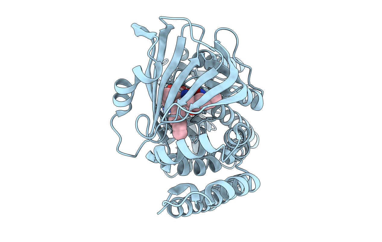
PDB ID:
3ATQ
Keywords:
Title:
Geranylgeranyl Reductase (GGR) from Sulfolobus acidocaldarius
Biological Source:
Source Organism(s):
Sulfolobus acidocaldarius (Taxon ID: 2285)
Expression System(s):
Method Details:
Experimental Method:
Resolution:
1.85 Å
R-Value Free:
0.20
R-Value Work:
0.18
R-Value Observed:
0.18
Space Group:
P 21 21 21
