Deposition Date
2011-01-04
Release Date
2012-01-18
Last Version Date
2024-03-13
Method Details:
Experimental Method:
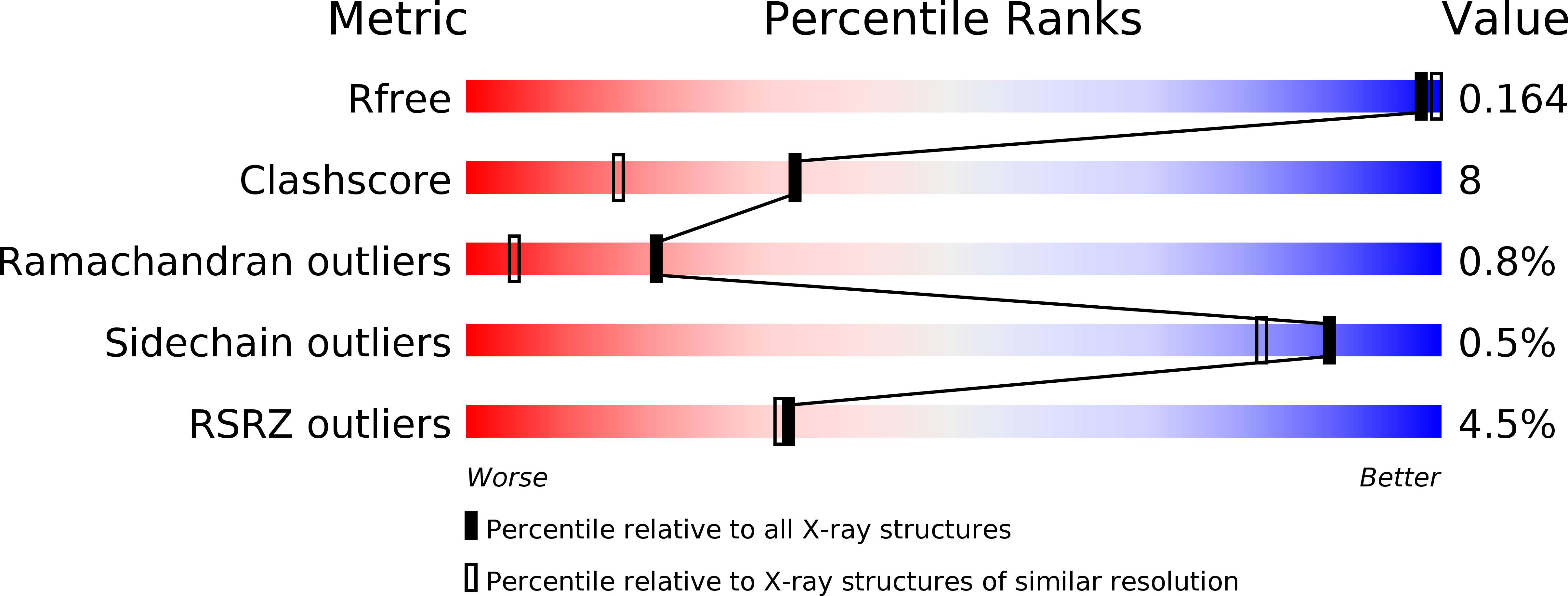
Resolution:
1.66 Å
R-Value Free:
0.16
R-Value Work:
0.14
R-Value Observed:
0.14
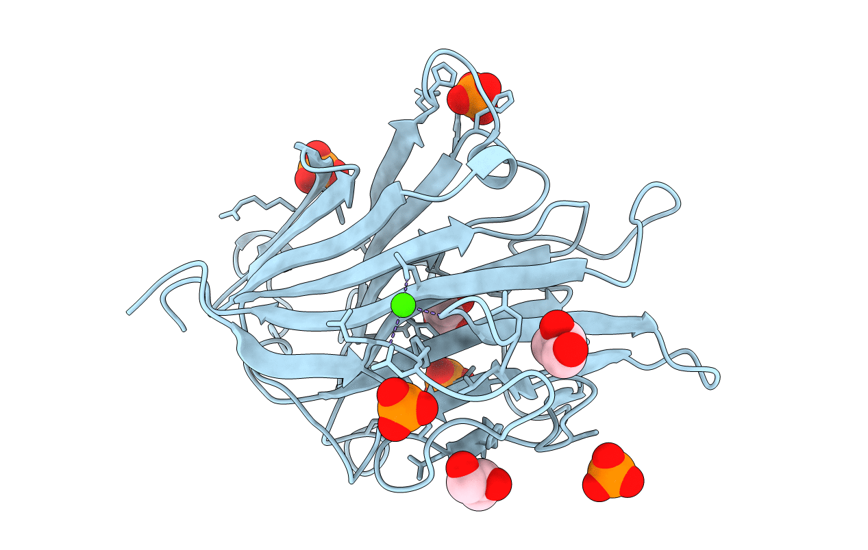
Space Group:
P 43