Deposition Date
2010-12-26
Release Date
2011-04-20
Last Version Date
2024-03-13
Entry Detail
PDB ID:
3AT5
Keywords:
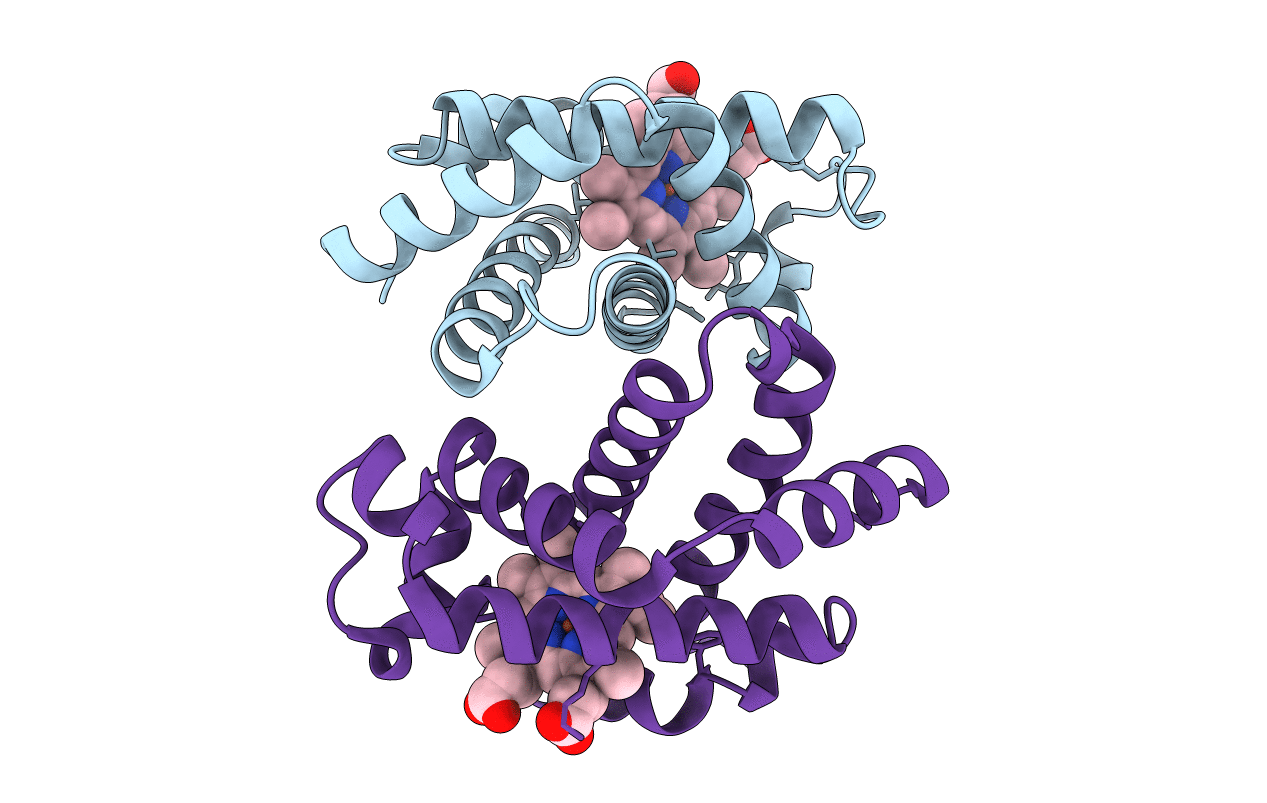
Title:
Side-necked turtle (Pleurodira, Chelonia, REPTILIA) hemoglobin: cDNA-derived primary structures and X-ray crystal structures of Hb A
Biological Source:
Source Organism(s):
Podocnemis unifilis (Taxon ID: 227871)
Method Details:
Experimental Method:
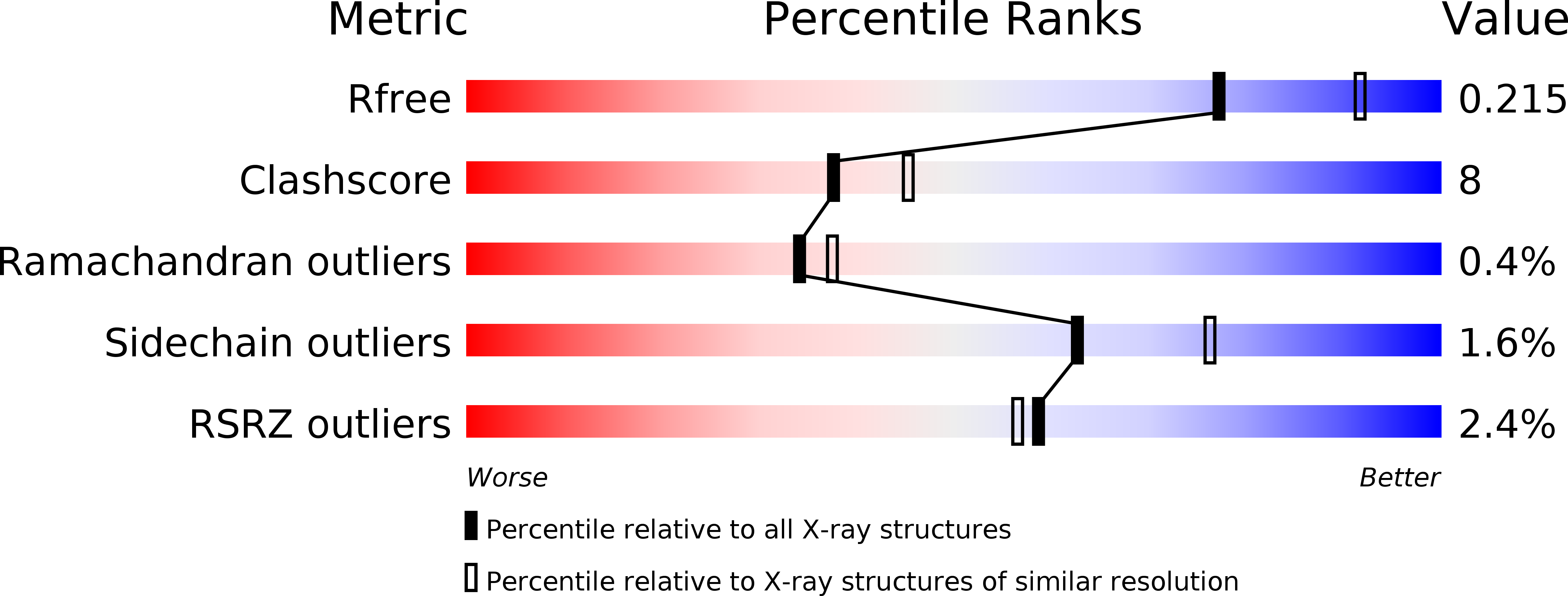
Resolution:
2.20 Å
R-Value Free:
0.22
R-Value Work:
0.20
Space Group:
P 41 21 2