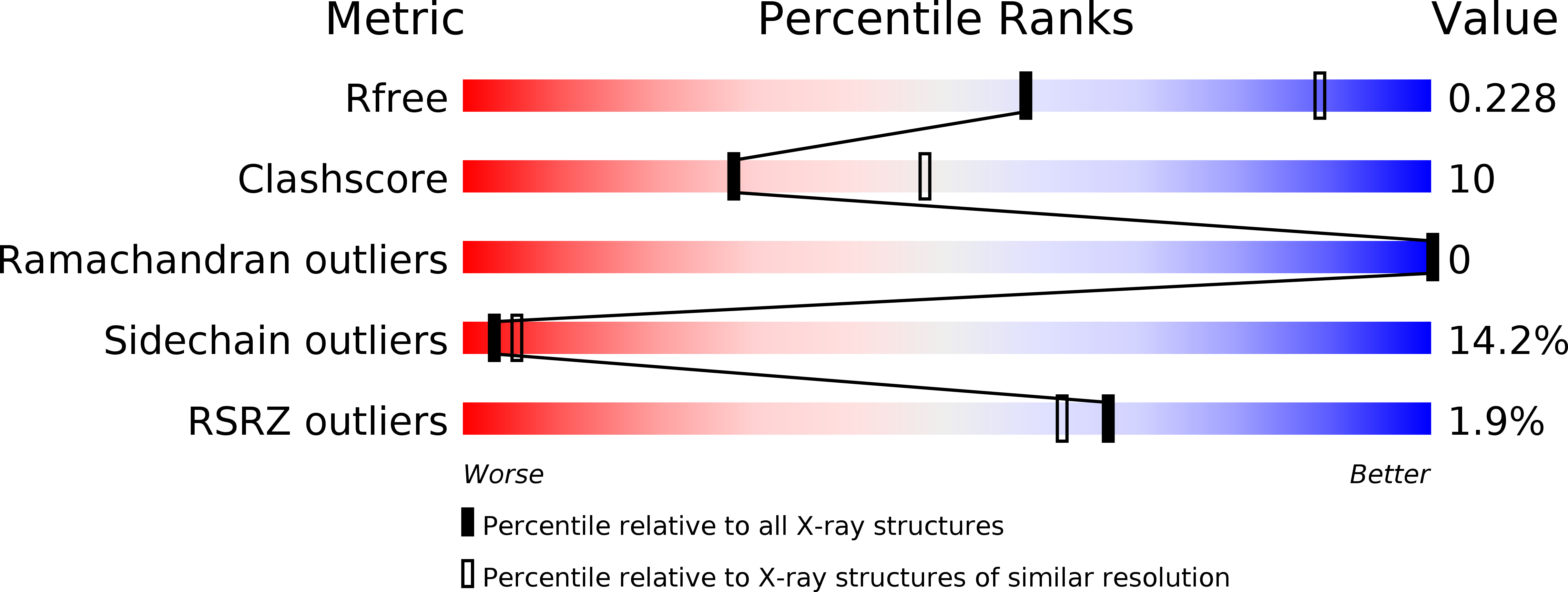
3ASM image
Deposition Date 2010-12-16
Release Date 2011-06-08
Last Version Date 2023-11-01
Entry Detail
PDB ID:
3ASM
Keywords:
Title:
Crystal structure of Q54A mutant protein of Bst-RNase HIII
Biological Source:
Source Organism:
Host Organism:
Method Details:
Experimental Method:
Resolution:
2.60 Å
R-Value Free:
0.23
R-Value Work:
0.20
R-Value Observed:
0.20
Space Group:
P 21 21 2
Macromolecular Entities
Structures with similar UniProt ID
Protein Blast
Polymer Type:polypeptide(L)
Molecule:Ribonuclease HIII
Gene (Uniprot):rnhC
Mutagens:Q54A
Chain IDs:A
Chain Length:310
Number of Molecules:1
Biological Source:Geobacillus stearothermophilus
Primary Citation
Identification of the substrate binding site in the N-terminal TBP-like domain of RNase H3.
Febs Lett. 585 2313 2317 (2011)
PMID: 21664908 DOI: 10.1016/j.febslet.2011.05.064

Abstact

Ribonuclease H3 from Bacillus stearothermophilus (Bst-RNase H3) has the N-terminal TBP-like substrate-binding domain. To identify the substrate binding site in this domain, the mutant proteins of the intact protein and isolated N-domain, in which six of the seventeen residues corresponding to those involved in DNA binding of TBP are individually mutated to Ala, were constructed. All of them exhibited decreased enzymatic activities and/or substrate-binding affinities when compared to those of the parent proteins, suggesting that the N-terminal domain of RNase H3 uses the flat surface of the β-sheet for substrate binding as TBP to bind DNA. This domain may greatly change conformation upon substrate binding.

Legend

Protein

Chemical

Disease

Primary Citation of related structures