3APQ image
Entry Detail
PDB ID:
3APQ
Keywords:
Title:
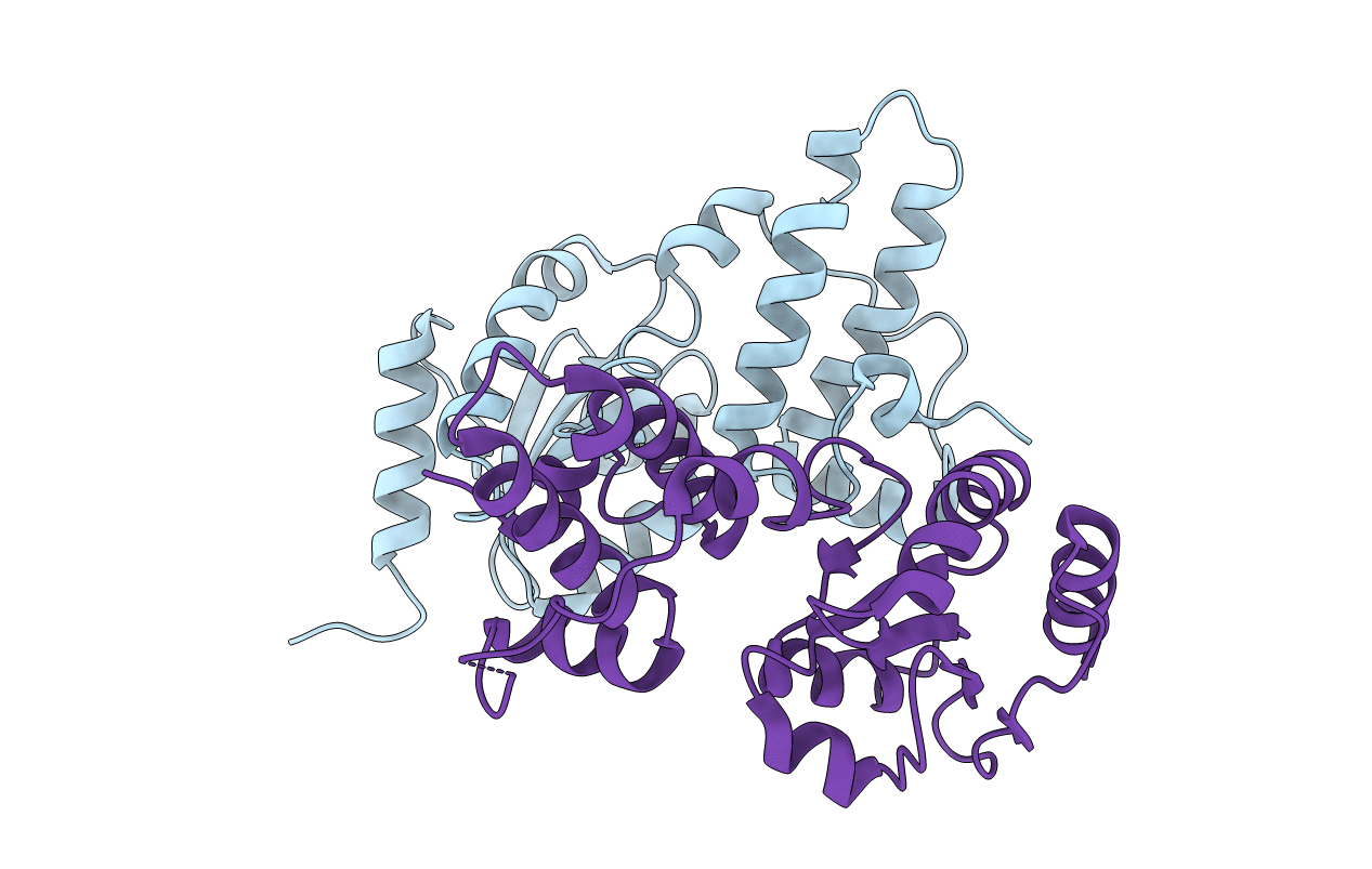
Crystal structure of J-Trx1 fragment of ERdj5
Biological Source:
Source Organism:
Host Organism:
PDB Version:
Deposition Date:
2010-10-20
Release Date:
2011-04-20
Method Details:
Experimental Method:
Resolution:
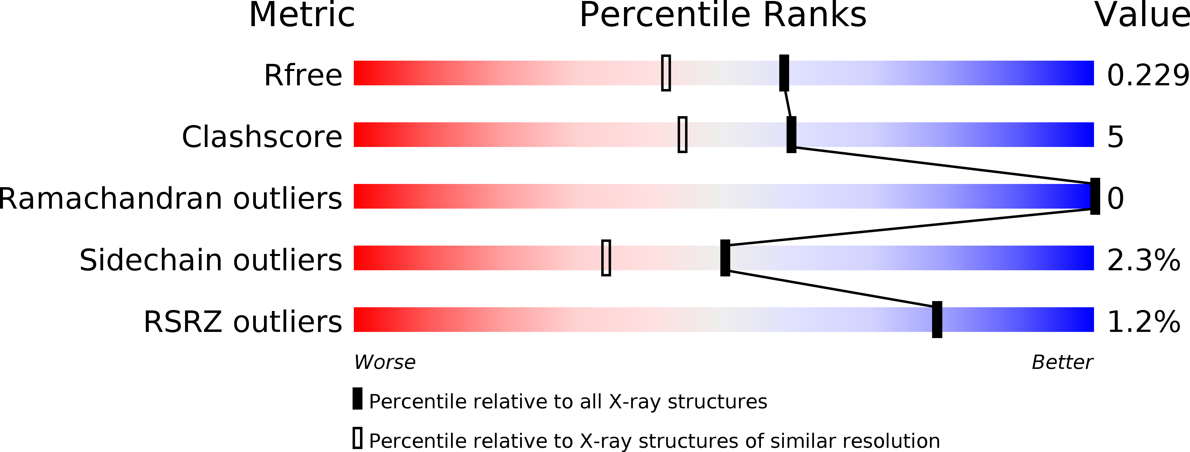
1.84 Å
R-Value Free:
0.23
R-Value Work:
0.17
R-Value Observed:
0.17
Space Group:
P 1 21 1
Macromolecular Entities
Polymer Type:polypeptide(L)
Description:DnaJ homolog subfamily C member 10
Chain IDs:A, B
Chain Length:210
Number of Molecules:2
Biological Source:Mus musculus
Primary Citation
Structural basis of an ERAD pathway mediated by the ER-resident protein disulfide reductase ERdj5.
Mol.Cell 41 432 444 (2011)
PMID: 21329881 DOI: 10.1016/j.molcel.2011.01.021

Abstact

ER-associated degradation (ERAD) is an ER quality-control process that eliminates terminally misfolded proteins. ERdj5 was recently discovered to be a key ER-resident PDI family member protein that accelerates ERAD by reducing incorrect disulfide bonds in misfolded glycoproteins recognized by EDEM1. We here solved the crystal structure of full-length ERdj5, thereby revealing that ERdj5 contains the N-terminal J domain and six tandem thioredoxin domains that can be divided into the N- and C-terminal clusters. Our systematic biochemical analyses indicated that two thioredoxin domains that constitute the C-terminal cluster form the highly reducing platform that interacts with EDEM1 and reduces EDEM1-recruited substrates, leading to their facilitated degradation. The pulse-chase experiment further provided direct evidence for the sequential movement of an ERAD substrate from calnexin to the downstream EDEM1-ERdj5 complex, and then to the retrotranslocation channel, probably through BiP. We present a detailed molecular view of how ERdj5 mediates ERAD in concert with EDEM1.

Legend

Protein

Chemical

Disease

Primary Citation of related structures