3AMC image
Deposition Date 2010-08-19
Release Date 2011-08-10
Last Version Date 2024-03-13
Entry Detail
PDB ID:
3AMC
Keywords:
Title:
Crystal structures of Thermotoga maritima Cel5A, apo form and dimer/au
Biological Source:
Source Organism:
Host Organism:
Method Details:
Experimental Method:
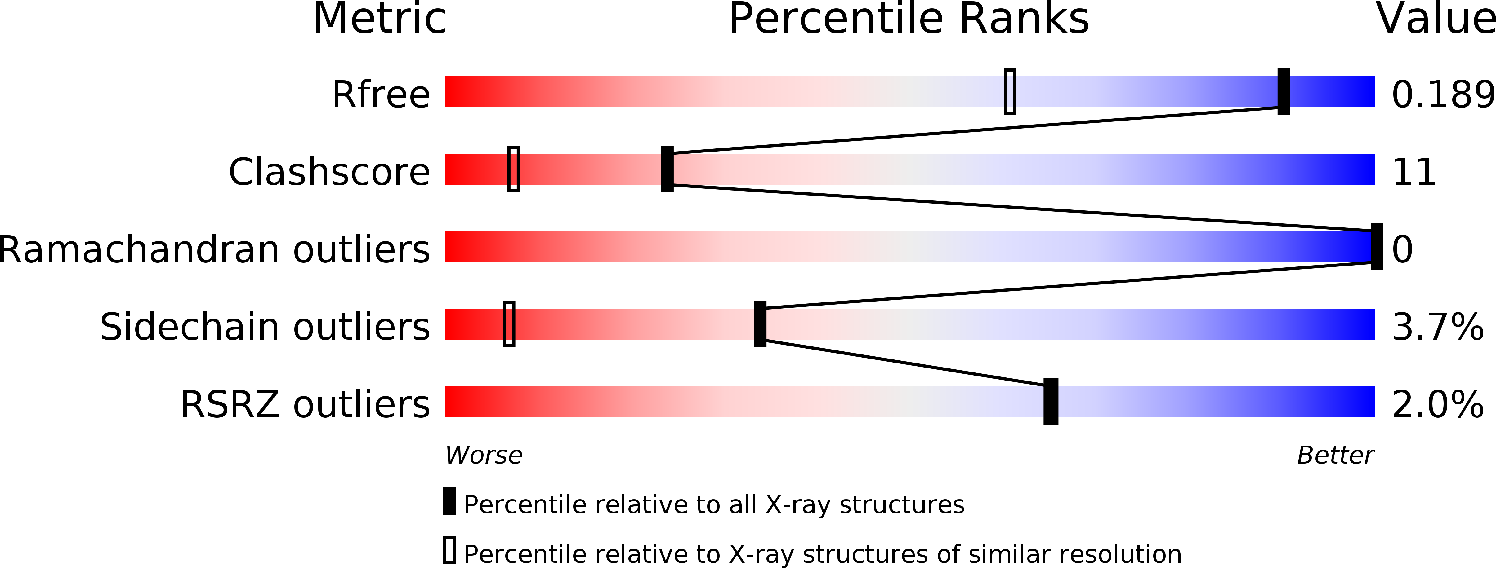
Resolution:
1.40 Å
R-Value Free:
0.20
R-Value Work:
0.18
Space Group:
P 1 21 1
Macromolecular Entities
Structures with similar UniProt ID
Protein Blast
Polymer Type:polypeptide(L)
Molecule:Endoglucanase
Gene (Uniprot):TM_1751
Chain IDs:A, B
Chain Length:317
Number of Molecules:2
Biological Source:Thermotoga maritima
Primary Citation
Diverse substrate recognition mechanism revealed by Thermotoga maritima Cel5A structures in complex with cellotetraose, cellobiose and mannotriose
Biochim.Biophys.Acta 1814 1832 1840 (2011)
PMID: 21839861 DOI: 10.1016/j.bbapap.2011.07.020

Abstact

The hyperthermophilic endoglucanase Cel5A from Thermotoga maritima can find applications in lignocellulosic biofuel production, because it catalyzes the hydrolysis of glucan- and mannan-based polysaccharides. Here, we report the crystal structures in apo-form and in complex with three ligands, cellotetraose, cellobiose and mannotriose, at 1.29Å to 2.40Å resolution. The open carbohydrate-binding cavity which can accommodate oligosaccharide substrates with extensively branched chains explained the dual specificity of the enzyme. Combining our structural information and the previous kinetic data, it is suggested that this enzyme prefers β-glucosyl and β-mannosyl moieties at the reducing end and uses two conserved catalytic residues, E253 (nucleophile) and E136 (general acid/base), to hydrolyze the glycosidic bonds. Moreover, our results also suggest that the wide spectrum of Tm_Cel5A substrates might be due to the lack of steric hindrance around the C2-hydroxyl group of the glucose or mannose unit from active-site residues.

Legend

Protein

Chemical

Disease

Primary Citation of related structures