3AM7 image
Deposition Date 2010-08-17
Release Date 2011-08-17
Last Version Date 2024-05-29
Entry Detail
PDB ID:
3AM7
Title:
Crystal structure of the ternary complex of eIF4E-M7GTP-4EBP2 peptide
Biological Source:
Source Organism:
Homo sapiens (Taxon ID: 9606)
Host Organism:
Method Details:
Experimental Method:
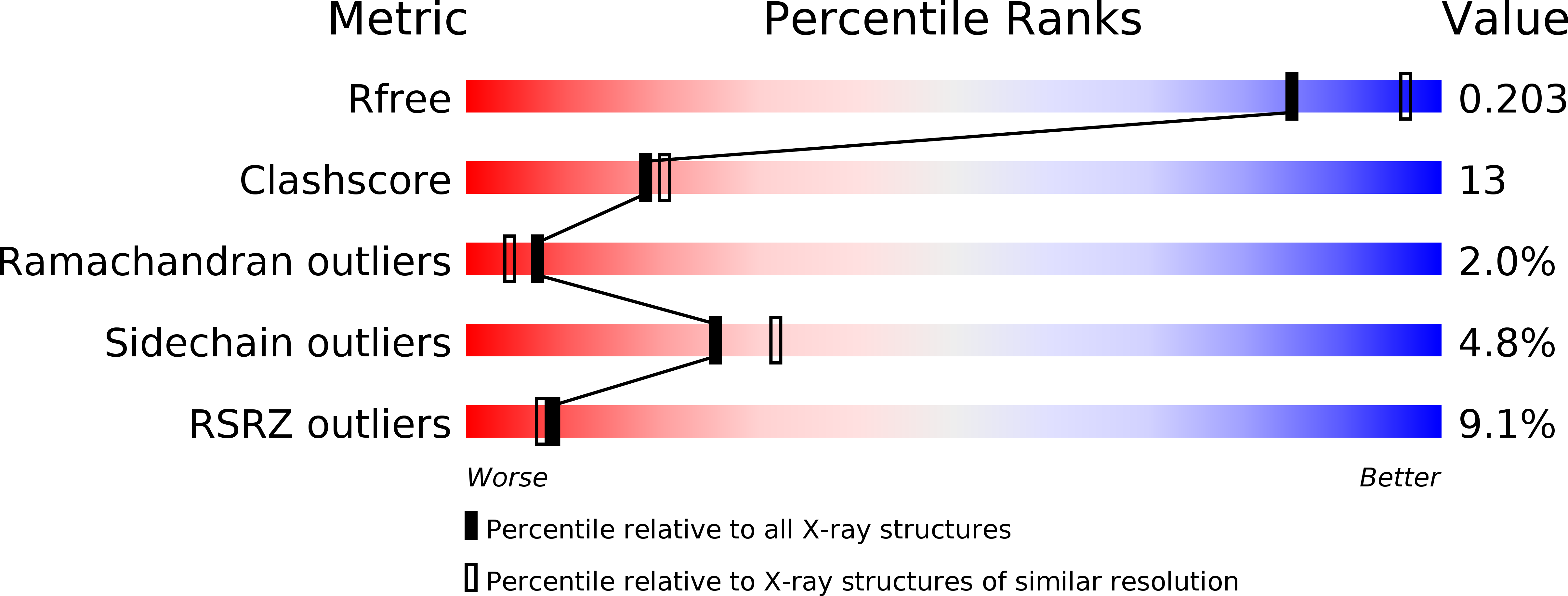
Resolution:
2.20 Å
R-Value Free:
0.26
R-Value Work:
0.23
Space Group:
P 43
Macromolecular Entities
Polymer Type:polypeptide(L)
Molecule:Eukaryotic translation initiation factor 4E
Gene (Uniprot):EIF4E
Chain IDs:A
Chain Length:191
Number of Molecules:1
Biological Source:Homo sapiens
Polymer Type:polypeptide(L)
Molecule:Eukaryotic translation initiation factor 4E-binding protein 2
Gene (Uniprot):EIF4EBP2
Chain IDs:B
Chain Length:19
Number of Molecules:1
Biological Source:Homo sapiens
Ligand Molecules
Primary Citation
Structural scaffold for eIF4E binding selectivity of 4E-BP isoforms: crystal structure of eIF4E binding region of 4E-BP2 and its comparison with that of 4E-BP1.
J.Pept.Sci. 17 650 657 (2011)
PMID: 21661078 DOI: 10.1002/psc.1384

Abstact

To clarify the higher eukaryotic initiation factor 4E (eIF4E) binding selectivity of 4E-binding protein 2 (4E-BP2) than of 4E-BP1, as determined by Trp fluorescence analysis, the crystal structure of the eIF4E binding region of 4E-BP2 in complex with m(7) GTP-bound human eIF4E has been determined by X-ray diffraction analysis and compared with that of 4E-BP1. The crystal structure revealed that the Pro47-Ser65 moiety of 4E-BP2 adopts a L-shaped conformation involving extended and α-helical structures and extends over the N-terminal loop and two different helix regions of eIF4E through hydrogen bonds, and electrostatic and hydrophobic interactions; these features were similarly observed for 4E-BP1. Although the pattern of the overall interaction of 4E-BP2 with eIF4E was similar to that of 4E-BP1, a notable difference was observed for the 60-63 sequence in relation to the conformation and binding selectivity of the 4E-BP isoform, i.e. Met-Glu-Cys-Arg for 4E-BP1 and Leu-Asp-Arg-Arg for 4E-BP2. In this paper, we report that the structural scaffold of the eIF4E binding preference for 4E-BP2 over 4E-BP1 is based on the stacking of the Arg63 planar side chain on the Trp73 indole ring of eIF4E and the construction of a compact hydrophobic space around the Trp73 indole ring by the Leu59-Leu60 sequence of 4E-BP2.

Legend

Protein

Chemical

Disease

Primary Citation of related structures