3AKI image
Deposition Date 2010-07-14
Release Date 2010-08-25
Last Version Date 2023-11-01
Entry Detail
PDB ID:
3AKI
Keywords:
Title:
Crystal structure of exo-1,5-alpha-L-arabinofuranosidase complexed with alpha-L-arabinofuranosyl azido
Biological Source:
Source Organism:
Host Organism:
Method Details:
Experimental Method:
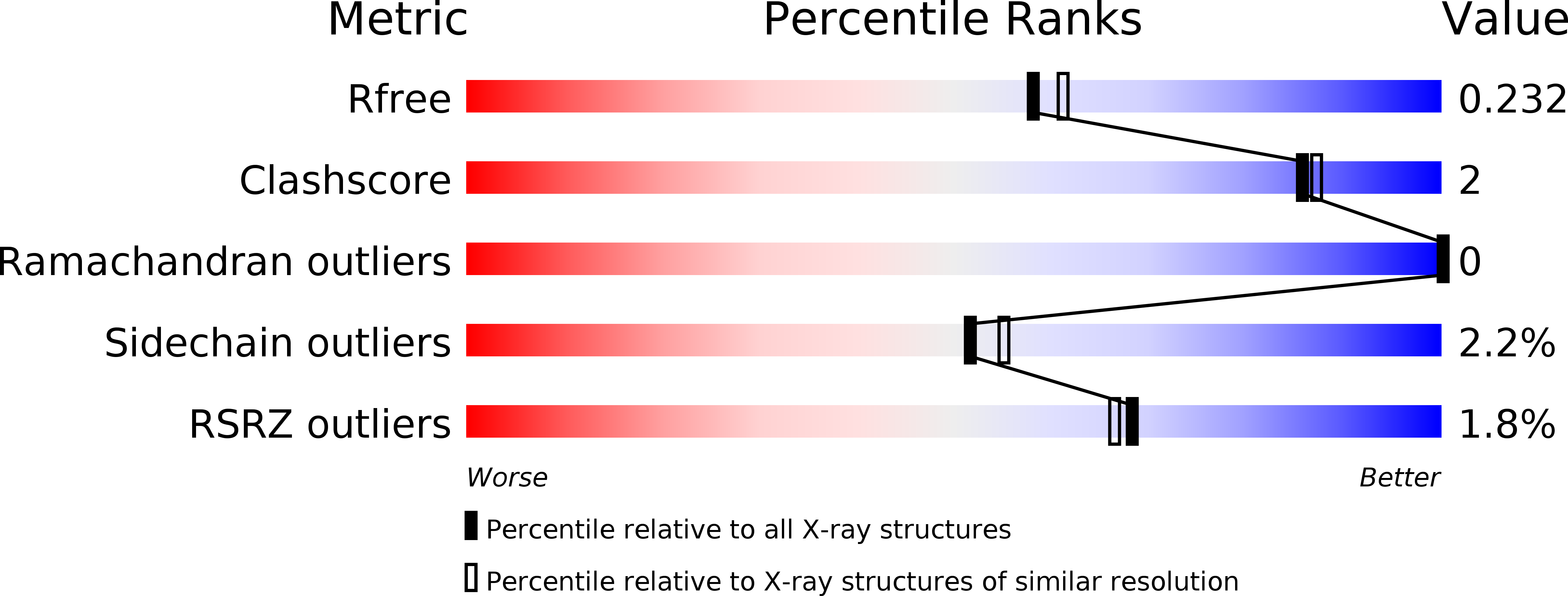
Resolution:
2.00 Å
R-Value Free:
0.23
R-Value Work:
0.19
R-Value Observed:
0.19
Space Group:
P 21 21 21
Macromolecular Entities
Structures with similar UniProt ID
Protein Blast
Polymer Type:polypeptide(L)
Molecule:Putative secreted alpha L-arabinofuranosidase II
Gene (Uniprot):Araf43A
Chain IDs:A
Chain Length:468
Number of Molecules:1
Biological Source:Streptomyces avermitilis
Primary Citation
Crystal Structure of an Exo-1,5-{alpha}-L-arabinofuranosidase from Streptomyces avermitilis Provides Insights into the Mechanism of Substrate Discrimination between Exo- and Endo-type Enzymes in Glycoside Hydrolase Family 43.
J.Biol.Chem. 285 34134 34143 (2010)
PMID: 20739278 DOI: 10.1074/jbc.M110.164251

Abstact

Exo-1,5-α-L-arabinofuranosidases belonging to glycoside hydrolase family 43 have strict substrate specificity. These enzymes hydrolyze only the α-1,5-linkages of linear arabinan and arabino-oligosaccharides in an exo-acting manner. The enzyme from Streptomyces avermitilis contains a core catalytic domain belonging to glycoside hydrolase family 43 and a C-terminal arabinan binding module belonging to carbohydrate binding module family 42. We determined the crystal structure of intact exo-1,5-α-L-arabinofuranosidase. The catalytic module is composed of a 5-bladed β-propeller topologically identical to the other family 43 enzymes. The arabinan binding module had three similar subdomains assembled against one another around a pseudo-3-fold axis, forming a β-trefoil-fold. A sugar complex structure with α-1,5-L-arabinofuranotriose revealed three subsites in the catalytic domain, and a sugar complex structure with α-L-arabinofuranosyl azide revealed three arabinose-binding sites in the carbohydrate binding module. A mutagenesis study revealed that substrate specificity was regulated by residues Asn-159, Tyr-192, and Leu-289 located at the aglycon side of the substrate-binding pocket. The exo-acting manner of the enzyme was attributed to the strict pocket structure of subsite -1, formed by the flexible loop region Tyr-281-Arg-294 and the side chain of Tyr-40, which occupied the positions corresponding to the catalytic glycon cleft of GH43 endo-acting enzymes.

Legend

Protein

Chemical

Disease

Primary Citation of related structures
Feedback Form
Name
Email
Institute
Feedback