Deposition Date
2010-06-29
Release Date
2011-05-18
Last Version Date
2023-11-01
Entry Detail
PDB ID:
3AK0
Keywords:
Title:
Crystal Structure of Ancestral Congerin Con-anc'-N28K
Method Details:
Experimental Method:
Resolution:
1.59 Å
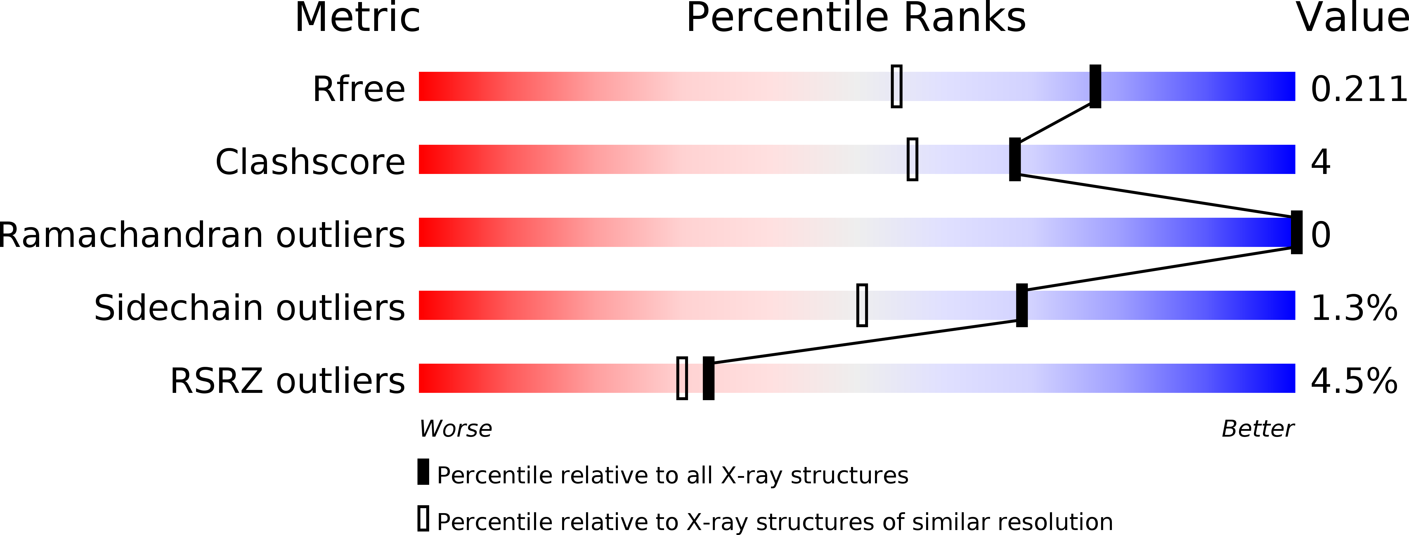
R-Value Free:
0.21
R-Value Work:
0.18
R-Value Observed:
0.18
Space Group:
P 21 21 2