3AJH image
Deposition Date 2010-06-05
Release Date 2011-03-16
Last Version Date 2024-03-13
Entry Detail
PDB ID:
3AJH
Keywords:
Title:
Crystal structure of PcyA V225D-biliverdin XIII alpha complex
Biological Source:
Source Organism:
Synechocystis (Taxon ID: 1148)
Host Organism:
Method Details:
Experimental Method:
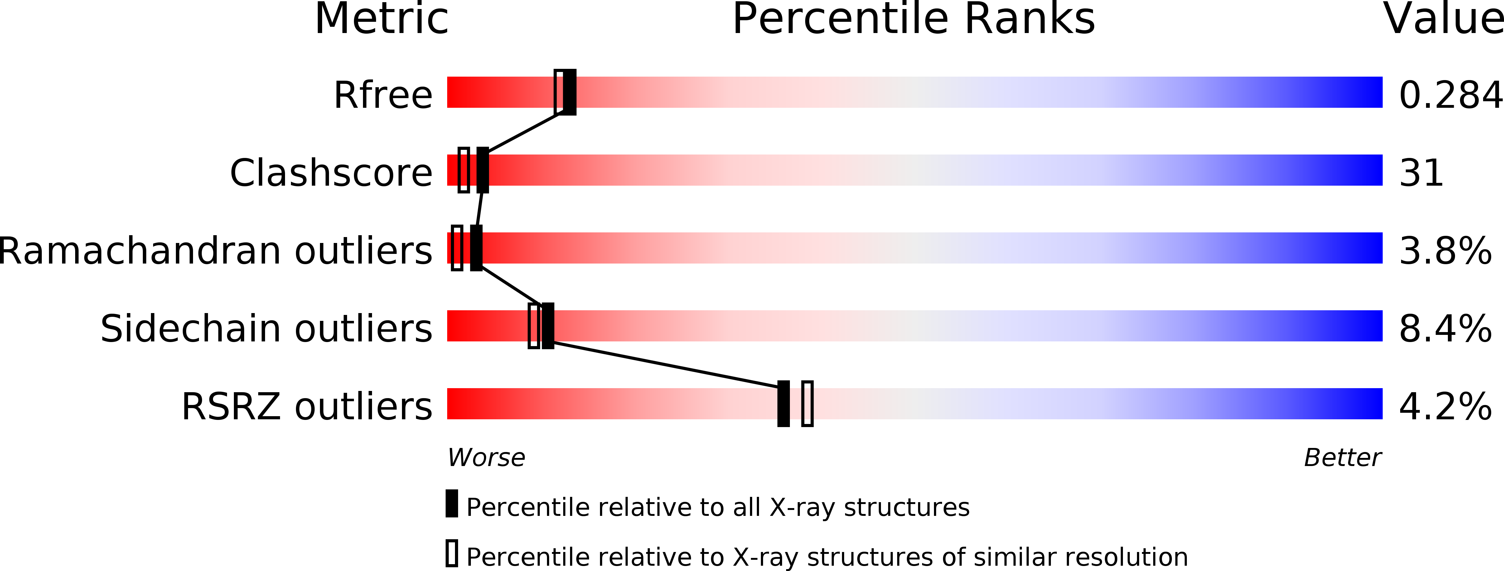
Resolution:
2.25 Å
R-Value Free:
0.28
R-Value Work:
0.25
R-Value Observed:
0.25
Space Group:
P 21 21 21
Macromolecular Entities
Structures with similar UniProt ID
Protein Blast
Polymer Type:polypeptide(L)
Molecule:Phycocyanobilin:ferredoxin oxidoreductase
Gene (Uniprot):pcyA
Chain IDs:A, B
Chain Length:248
Number of Molecules:2
Biological Source:Synechocystis
Ligand Molecules
Primary Citation
One residue substitution in PcyA leads to unexpected changes in tetrapyrrole substrate binding.
Biochem.Biophys.Res.Commun. 402 373 377 (2010)
PMID: 20946883 DOI: 10.1016/j.bbrc.2010.10.037

Abstact

Phycocyanobilin:ferredoxin oxidoreductase (PcyA) catalyzes the sequential reduction of the vinyl group of the D-ring and A-ring of biliverdin IXα (BV), using reducing equivalents provided by ferredoxin. This reaction produces phycocyanobilin, a pigment used for light-harvesting and light-sensing in red algae and cyanobacteria. The crystal structure of PcyA-BV reveals that BV is specifically bound in the PcyA active pocket through extensive hydrophobic and hydrophilic interactions. During the course of a mutational study of PcyA, we observed that mutation of the V225 position, apart from the processing sites, conferred an unusual property on PcyA; V225D mutant protein could bind BV and its analog BV13, but these complexes showed a distinct UV-vis absorption spectrum from that of the wild-type PcyA-BV complex. The crystal structures of BV- and BV13-bound forms of V225D protein revealed that gross structural changes occurred near the substrate-binding pocket, and that the BV/BV13 binding manner in the pocket was dramatically altered. Protein folding in V225D-BV/BV13 was more similar to that of substrate-free PcyA than that in PcyA-BV; the "induced-fit" did not occur when BV/BV13 was bound to the V225D protein. The unexpected structural change presented here provides a cautionary note about interpreting functional data derived from a mutated protein in the absence of its exact structure.

Legend

Protein

Chemical

Disease

Primary Citation of related structures