3AIW image
Entry Detail
PDB ID:
3AIW
Keywords:
Title:
Crystal structure of beta-glucosidase in rye complexed with 2-deoxy-2-fluoroglucoside and dinitrophenol
Biological Source:
Source Organism:
Host Organism:
PDB Version:
Deposition Date:
2010-05-18
Release Date:
2011-02-23
Method Details:
Experimental Method:
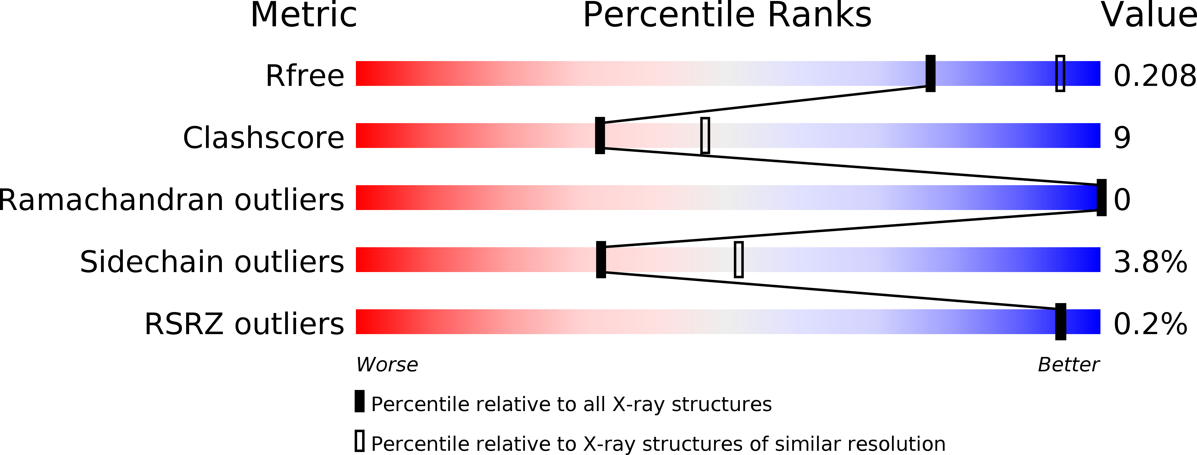
Resolution:
2.40 Å
R-Value Free:
0.21
R-Value Work:
0.18
R-Value Observed:
0.18
Space Group:
P 41 3 2
Macromolecular Entities
Polymer Type:polypeptide(L)
Description:Beta-glucosidase
Chain IDs:A
Chain Length:564
Number of Molecules:1
Biological Source:Secale cereale
Primary Citation
Active-site architecture of benzoxazinone-glucoside beta-D-glucosidases in Triticeae
Plant Sci. 180 268 275 (2011)
PMID: 21421370 DOI: 10.1016/j.plantsci.2010.09.001

Abstact

The β-D-glucosidases from wheat (Triticum aestivum) and rye (Secale cereale) hydrolyze benzoxazinone-glucose conjugates. Although wheat and rye glucosidases have high sequence identity, they have different substrate preferences; the wheat enzyme favors DIMBOA-Glc (2-O-β-D-glucopyranosyl-4-hydroxy-7-methoxy-1,4-benzoxazin-3-one) over DIBOA-Glc (7-demethoxy-DIMBOA-Glc), whereas the rye enzyme preference is the opposite. To investigate the mechanism of substrate binding, we analyzed crystal structures of an inactive mutant of the wheat glucosidase complexed with the natural substrate DIMBOA-Glc, wheat and rye glucosidases complexed with an aglycone DIMBOA, and wheat and rye glucosidases complexed with an inhibitor 2-fluoro-2-deoxy-β-D-glucose. The binding position of substrate in the active site was determined but interaction between the substrate and Ser-464 or Leu-465 was not observed, although amino acid residues at these two positions are the only structural distinctions between wheat and rye glucosidase catalytic pockets. Variation at these two positions alters the width of the pocket entrance, which may relate to observed differences in substrate specificity. The side chain of Glu-462 that forms hydrogen bonds with the glucose moiety of DIMBOA-Glc moved deeper into the pocket upon substrate binding, and mutation of this residue dramatically decreased enzyme activity.

Legend

Protein

Chemical

Disease

Primary Citation of related structures