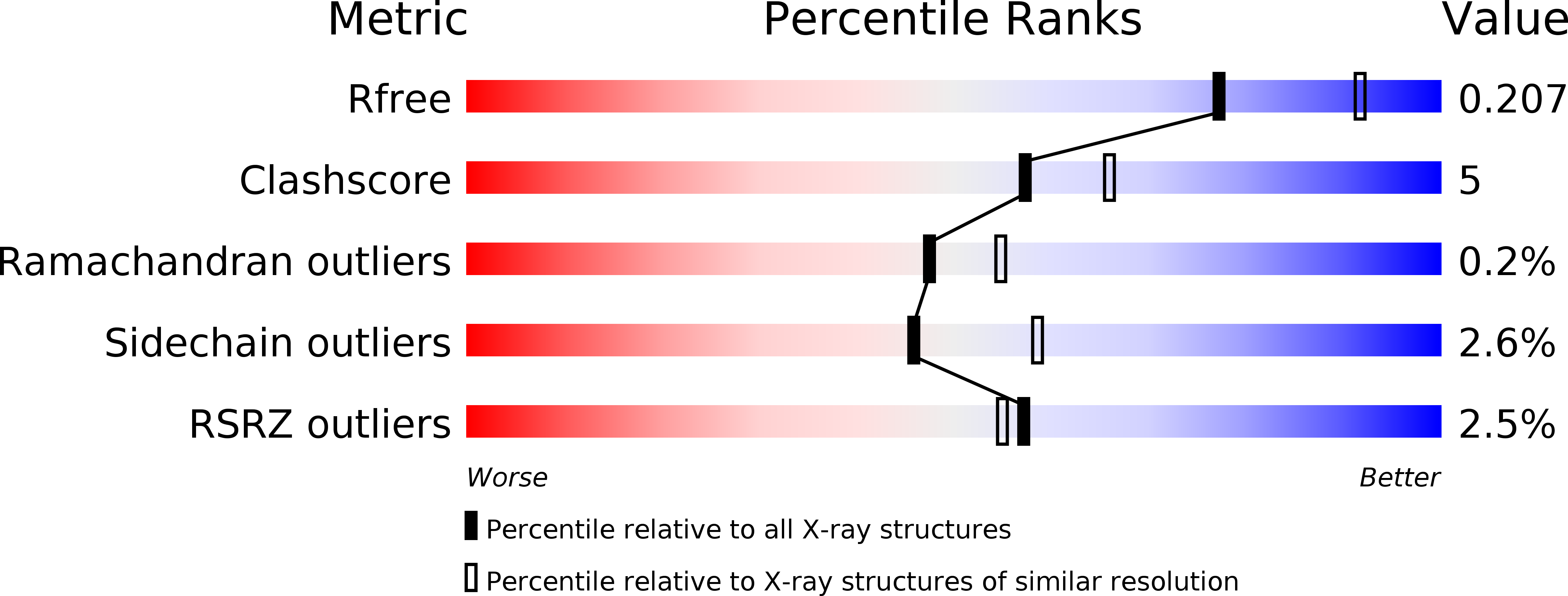
3AI7 image
Deposition Date 2010-05-10
Release Date 2010-09-15
Last Version Date 2024-04-03
Entry Detail
PDB ID:
3AI7
Keywords:
Title:
Crystal Structure of Bifidobacterium Longum Phosphoketolase
Biological Source:
Source Organism:
Host Organism:
Method Details:
Experimental Method:
Resolution:
2.20 Å
R-Value Free:
0.20
R-Value Work:
0.14
R-Value Observed:
0.14
Space Group:
P 1 21 1
Macromolecular Entities
Polymer Type:polypeptide(L)
Molecule:Xylulose-5-phosphate/fructose-6-phosphate phosphoketolase
Gene (Uniprot):xfp
Chain IDs:A, B, C, D, E, F, G, H
Chain Length:831
Number of Molecules:8
Biological Source:Bifidobacterium longum
Primary Citation
Crystal structure of Bifidobacterium Longum phosphoketolase; key enzyme for glucose metabolism in Bifidobacterium
Febs Lett. 584 3855 3861 (2010)
PMID: 20674574 DOI: 10.1016/j.febslet.2010.07.043

Abstact

The crystal structure of Bifidobacterium longum phosphoketolase, a thiamine diphosphate (TPP) dependent enzyme, has been determined at 2.2A resolution. The enzyme is a dimer with the active sites located at the interface between the two identical subunits with molecular mass of 92.5 kDa. The bound TPP is almost completely shielded from solvent except for the catalytically important C2-carbon of the thiazolium ring, which can be accessed by a substrate sugar through a narrow funnel-shaped channel. In silico docking studies of B. longum phosphoketolase with its substrate enable us to propose a model for substrate binding.

Legend

Protein

Chemical

Disease

Primary Citation of related structures