3AHW image
Deposition Date 2010-05-04
Release Date 2010-07-07
Last Version Date 2024-10-16
Entry Detail
PDB ID:
3AHW
Keywords:
Title:
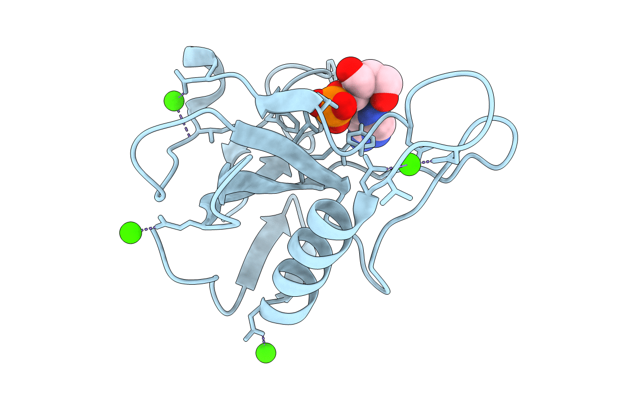
Crystal Structure of Ustilago sphaerogena Ribonuclease U2 complexed with adenosine 2'-monophosphate
Biological Source:
Source Organism:
Method Details:
Experimental Method:
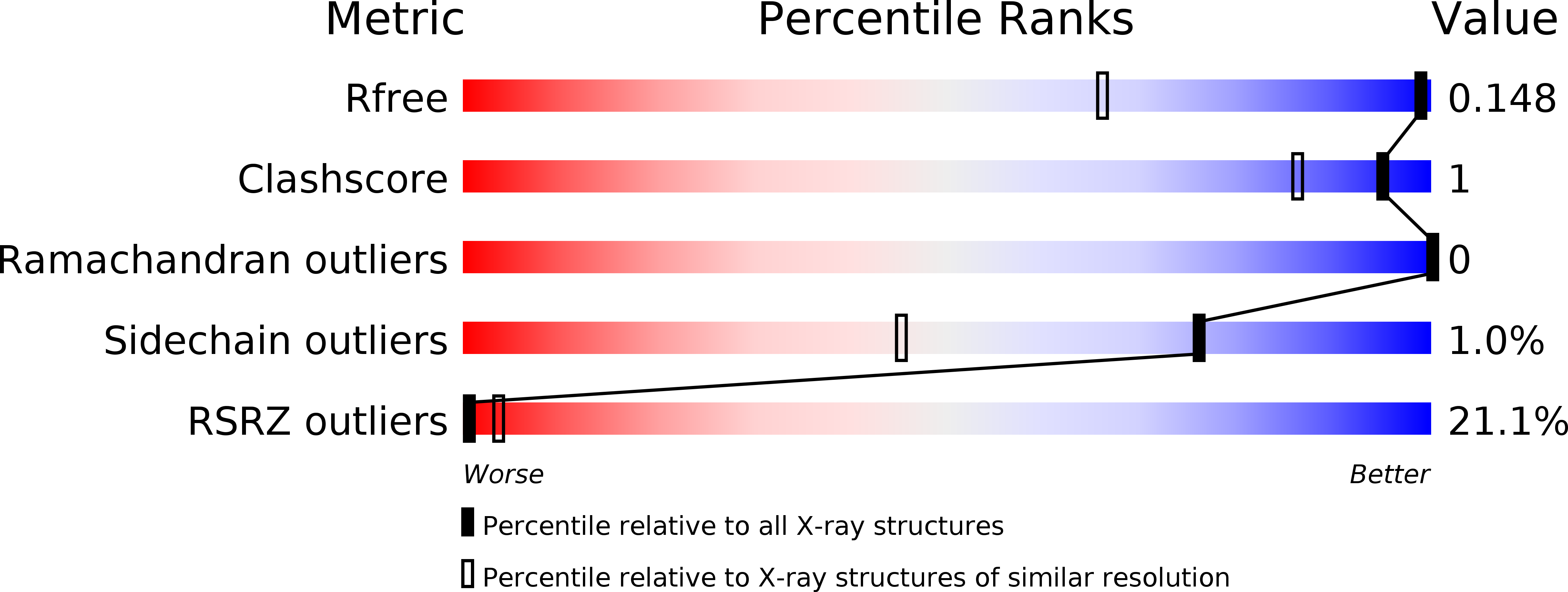
Resolution:
1.03 Å
R-Value Free:
0.14
R-Value Work:
0.13
R-Value Observed:
0.13
Space Group:
P 1 21 1
Macromolecular Entities
Structures with similar UniProt ID
Protein Blast
Polymer Type:polypeptide(L)
Molecule:Ribonuclease U2
Gene (Uniprot):RNU2
Chain IDs:A
Chain Length:114
Number of Molecules:1
Biological Source:Ustilago sphaerogena
Primary Citation
Conformational variation revealed by the crystal structure of RNase U2A complexed with Ca ion and 2'-adenylic acid at 1.03 angstrom resolution.
Protein Pept.Lett. 17 1559 1561 (2010)
PMID: 20858208 DOI: 10.2174/0929866511009011559

Abstact

Asparagine can be non-enzymatically deamidated and isomerized via succinimide to isoaspartate. This post-translational modification can potentially alter the physical properties or the function of the parent protein. Asn32 of ribonuclease U2A from Ustilago sphaerogena is known to rapidly deamidate and isomerize in alkaline conditions. The crystal structure of ribonuclease U2A complexed with 2'-adenylic acid and calcium ions was determined at 1.03 Å resolution. In this structure, the region from Asp29 to Asp37 winds around a calcium ion, and the main-chain of Asn32-Gly33 adopts an extended conformation. Rotation of the side-chain of Asn32 could bring Asn32C(γ) into close proximity to Gly33N, in a conformation suitable for succinimide formation. The structure suggests that in solution the region around Asn32-Gly33 is likely to be in equilibrium between multiple conformers, with the deamidation of Asn32 proceeding when the region adopts an extended conformation.

Legend

Protein

Chemical

Disease

Primary Citation of related structures
Feedback Form
Name
Email
Institute
Feedback