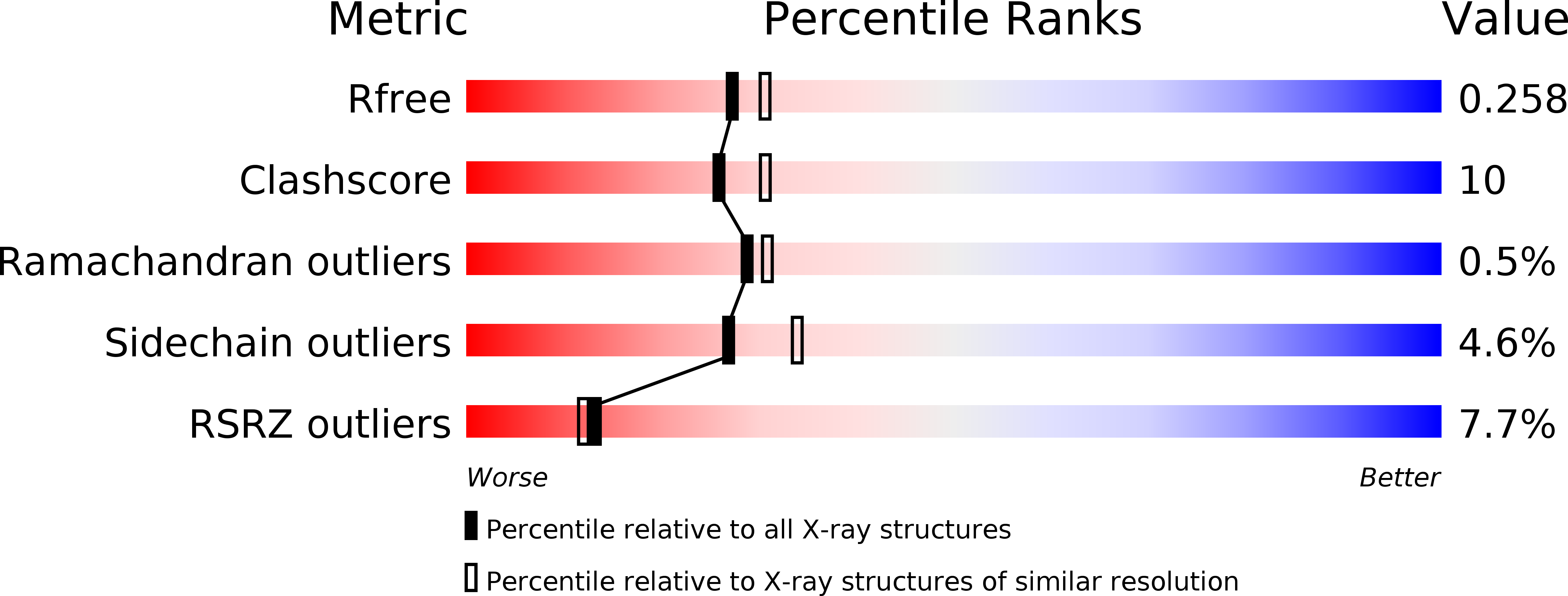
3AGW image
Deposition Date 2010-04-08
Release Date 2010-09-29
Last Version Date 2023-11-01
Entry Detail
PDB ID:
3AGW
Title:
Crystal Structure of the Cytoplasmic Domain of G-Protein-Gated Inward Rectifier Potassium Channel Kir3.2 in the absence of Na+
Biological Source:
Source Organism:
Mus musculus (Taxon ID: 10090)
Host Organism:
Method Details:
Experimental Method:
Resolution:
2.20 Å
R-Value Free:
0.23
R-Value Work:
0.19
R-Value Observed:
0.19
Space Group:
I 4 2 2
Macromolecular Entities
Structures with similar UniProt ID
Protein Blast
Polymer Type:polypeptide(L)
Molecule:G protein-activated inward rectifier potassium channel 2
Chain IDs:A
Chain Length:208
Number of Molecules:1
Biological Source:Mus musculus
Primary Citation
A structural determinant for the control of PIP2 sensitivity in G protein-gated inward rectifier K+ channels
J.Biol.Chem. 285 38517 38523 (2010)
PMID: 20880843 DOI: 10.1074/jbc.M110.161703

Abstact

Inward rectifier K(+) (Kir) channels are activated by phosphatidylinositol-(4,5)-bisphosphate (PIP(2)), but G protein-gated Kir (K(G)) channels further require either G protein βγ subunits (Gβγ) or intracellular Na(+) for their activation. To reveal the mechanism(s) underlying this regulation, we compared the crystal structures of the cytoplasmic domain of K(G) channel subunit Kir3.2 obtained in the presence and the absence of Na(+). The Na(+)-free Kir3.2, but not the Na(+)-plus Kir3.2, possessed an ionic bond connecting the N terminus and the CD loop of the C terminus. Functional analyses revealed that the ionic bond between His-69 on the N terminus and Asp-228 on the CD loop, which are known to be critically involved in Gβγ- and Na(+)-dependent activation, lowered PIP(2) sensitivity. The conservation of these residues within the K(G) channel family indicates that the ionic bond is a character that maintains the channels in a closed state by controlling the PIP(2) sensitivity.

Legend

Protein

Chemical

Disease

Primary Citation of related structures