3AFR image
Deposition Date 2010-03-10
Release Date 2010-03-31
Last Version Date 2023-11-01
Entry Detail
PDB ID:
3AFR
Title:
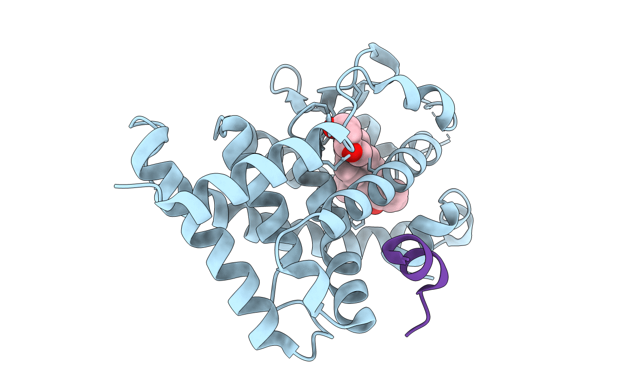
Crystal Structure of VDR-LBD/22S-Butyl-1a,24R-dihydroxyvitamin D3 complex
Biological Source:
Source Organism:
Host Organism:
Method Details:
Experimental Method:
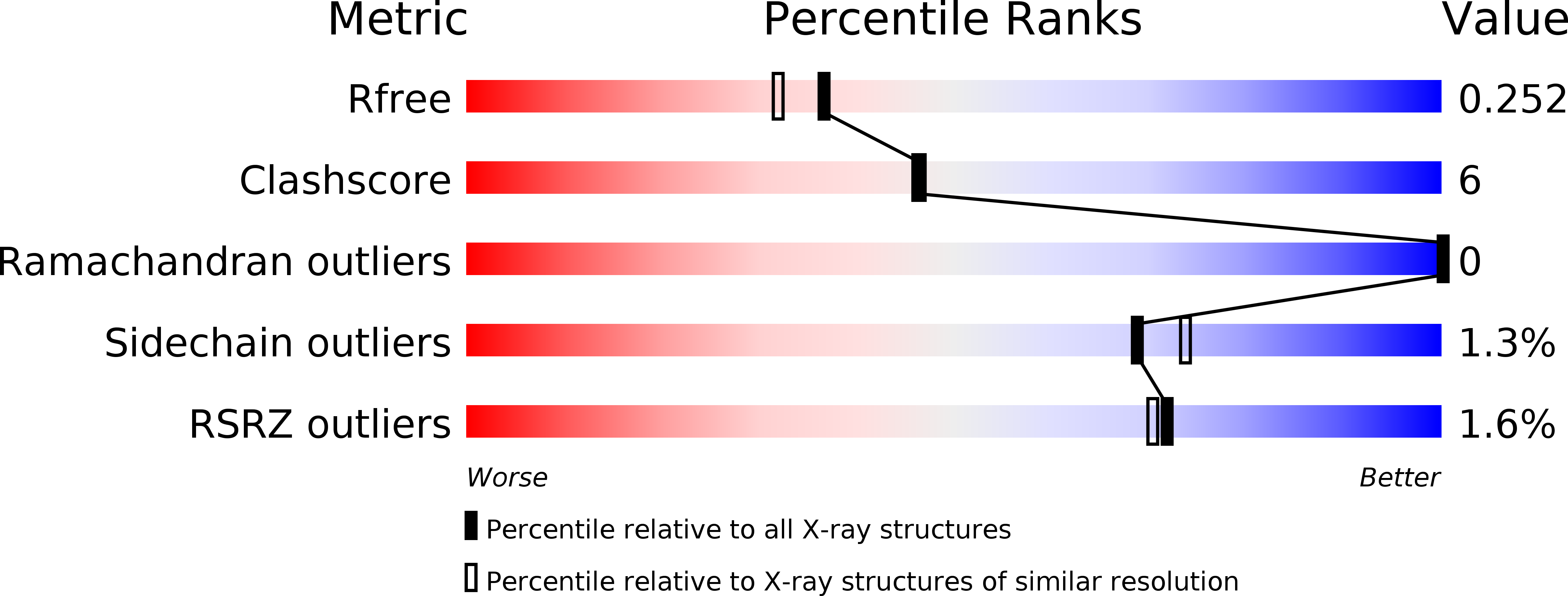
Resolution:
2.00 Å
R-Value Free:
0.26
R-Value Work:
0.21
Space Group:
C 1 2 1
Macromolecular Entities
Polymer Type:polypeptide(L)
Molecule:Vitamin D3 receptor
Gene (Uniprot):Vdr
Mutations:deletion mutant, residues 165-211
Chain IDs:A
Chain Length:271
Number of Molecules:1
Biological Source:Rattus norvegicus
Polymer Type:polypeptide(L)
Molecule:13-meric peptide from Mediator of RNA polymerase II transcription subunit 1
Gene (Uniprot):med1
Chain IDs:B (auth: C)
Chain Length:13
Number of Molecules:1
Biological Source:Xenopus tropicalis
Ligand Molecules
Primary Citation
22S-Butyl-1alpha,24R-dihydroxyvitamin D(3): Recovery of vitamin D receptor agonistic activity
J.Steroid Biochem.Mol.Biol. 121 146 150 (2010)
PMID: 20211257 DOI: 10.1016/j.jsbmb.2010.02.033

Abstact

We recently reported that 22S-butyl-1alpha,24R-dihydroxyvitamin D(3)3 recovers the agonistic activity for vitamin D receptor (VDR), although its 25,26,27-trinor analog 2 is a potent VDR antagonist. To investigate the structural features involved in the recovery of agonism, we crystallized the ternary complex of VDR-ligand-binding domain, ligand 3 and coactivator peptide, and conducted X-ray crystallographic analysis of the complex. Compared with the complex with 2, the complex with 3 recovered the following structural features: a pincer-type hydrogen bond between the 24-hydroxyl group and VDR, the conformation of Leu305, the positioning of His301 and His393, the stability of the complex, and intimate hydrophobic interactions between the ligand and helix 12. In addition, we evaluated the potency of both compounds for recruiting RXR and coactivator. The results indicate that the complex with 3 generates a suitable surface for coactivator recruitment. These studies suggest that the action of 2 as an antagonist is caused by the generation of a surface not suitable for coactivator recruitment due to the lack of hydrophobic interactions with helix 12 as well as insufficient hydrogen bond formation between the 24-hydroxyl group and VDR. We concluded that the action of 3 as an agonist is based on the elimination of these structural defects in the complex with 2.

Legend

Protein

Chemical

Disease

Primary Citation of related structures