Deposition Date
2010-01-22
Release Date
2010-05-26
Last Version Date
2023-11-01
Entry Detail
PDB ID:
3ADL
Keywords:
Title:
Structure of TRBP2 and its molecule implications for miRNA processing
Biological Source:
Source Organism(s):
Homo sapiens (Taxon ID: 9606)
Expression System(s):
Method Details:
Experimental Method:
Resolution:
2.20 Å
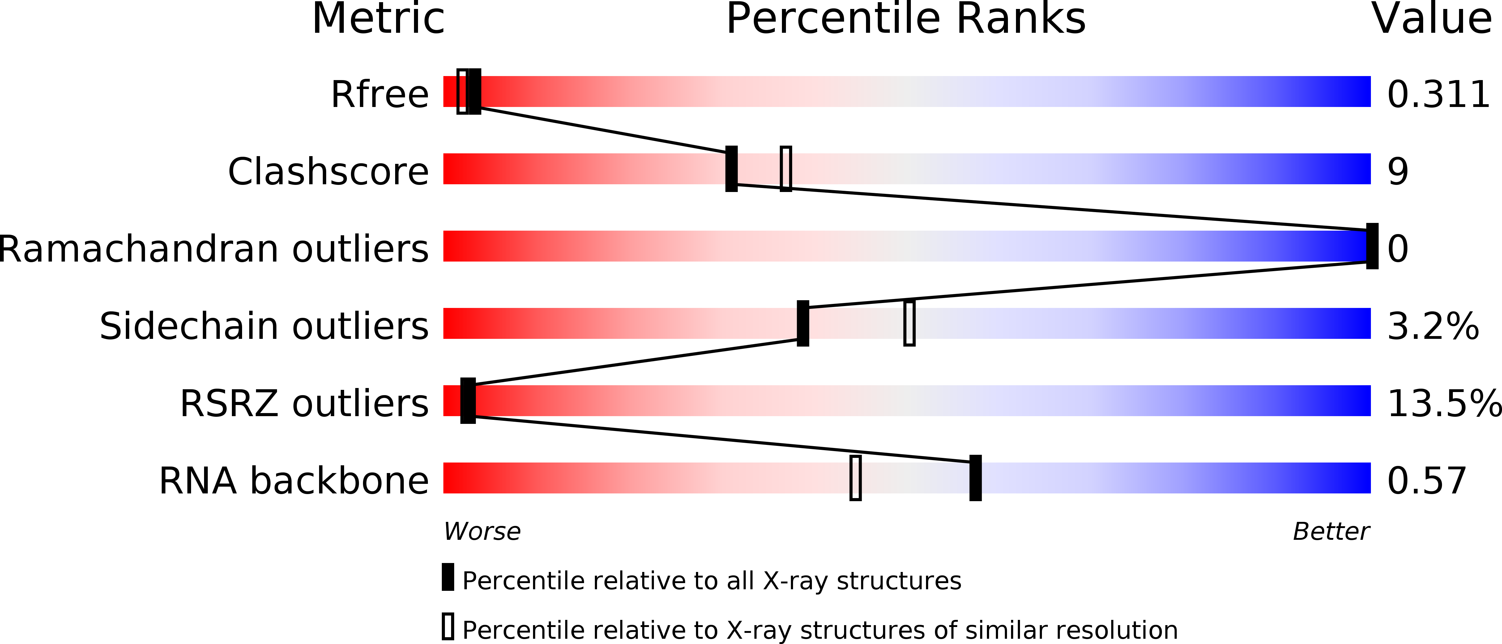
R-Value Free:
0.29
R-Value Work:
0.26
R-Value Observed:
0.26
Space Group:
I 21 21 21