3AD8 image
Deposition Date 2010-01-15
Release Date 2010-08-25
Last Version Date 2023-11-01
Entry Detail
PDB ID:
3AD8
Keywords:
Title:
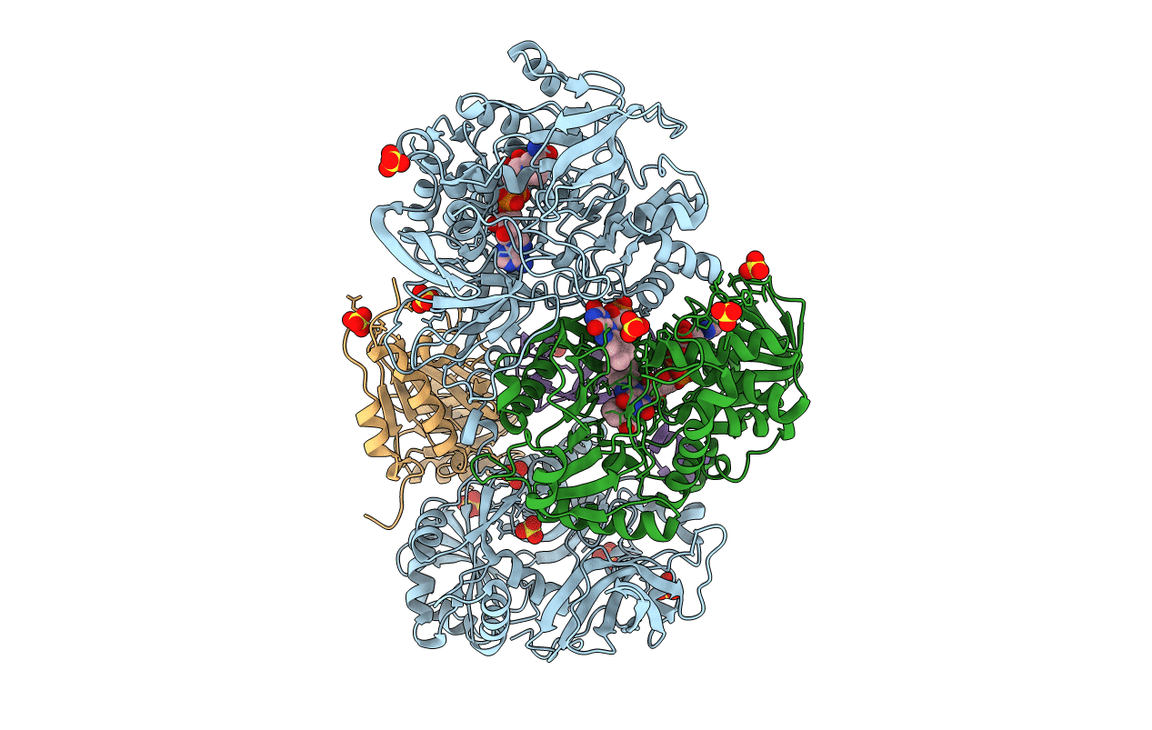
Heterotetrameric Sarcosine Oxidase from Corynebacterium sp. U-96 in complex with pyrrole 2-carboxylate
Biological Source:
Source Organism:
Host Organism:
Method Details:
Experimental Method:
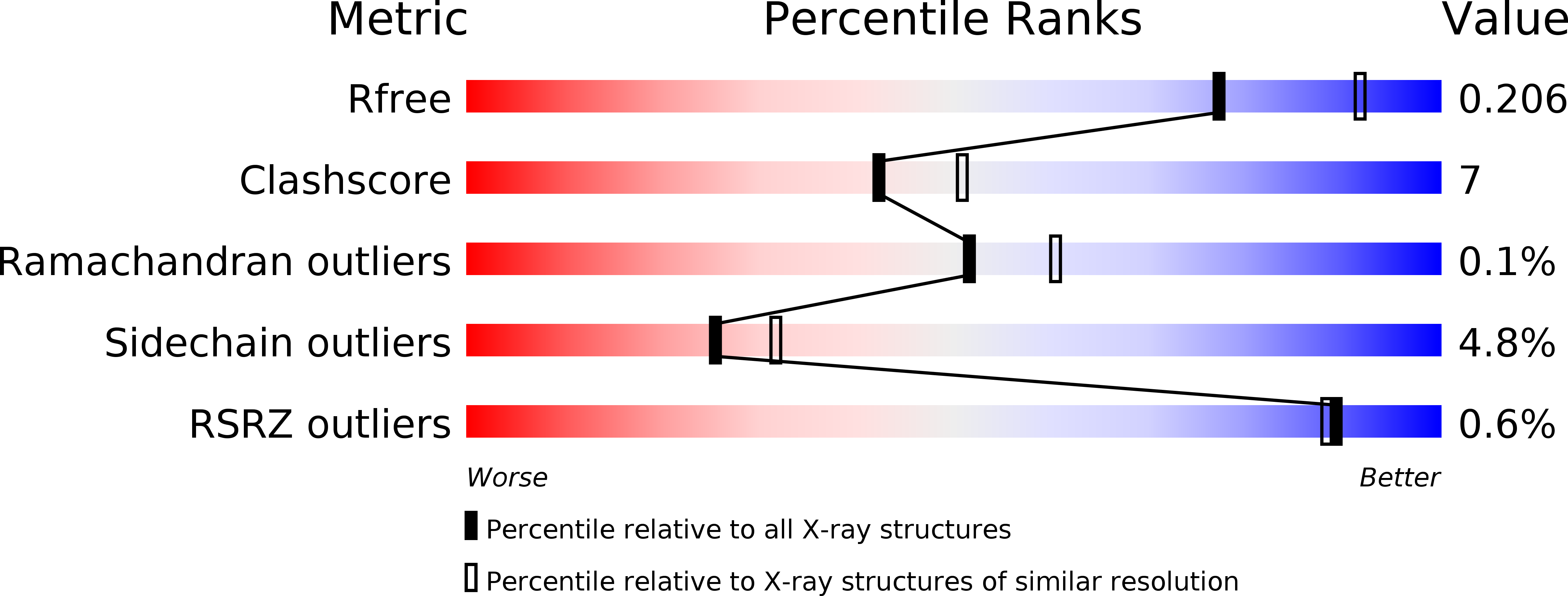
Resolution:
2.20 Å
R-Value Free:
0.20
R-Value Work:
0.16
R-Value Observed:
0.16
Space Group:
P 65 2 2
Macromolecular Entities
Polymer Type:polypeptide(L)
Molecule:SARCOSINE OXIDASE ALPHA SUBUNIT
Gene (Uniprot):soxA
Chain IDs:A
Chain Length:964
Number of Molecules:1
Biological Source:Corynebacterium sp. U-96
Polymer Type:polypeptide(L)
Molecule:SARCOSINE OXIDASE BETA SUBUNIT
Gene (Uniprot):soxB
Chain IDs:B
Chain Length:404
Number of Molecules:1
Biological Source:Corynebacterium sp. U-96
Polymer Type:polypeptide(L)
Molecule:SARCOSINE OXIDASE GAMMA SUBUNIT
Gene (Uniprot):soxG
Chain IDs:C
Chain Length:203
Number of Molecules:1
Biological Source:Corynebacterium sp. U-96
Polymer Type:polypeptide(L)
Molecule:SARCOSINE OXIDASE DELTA SUBUNIT
Gene (Uniprot):soxD
Chain IDs:D
Chain Length:99
Number of Molecules:1
Biological Source:Corynebacterium sp. U-96
Primary Citation
Channeling and conformational changes in the heterotetrameric sarcosine oxidase from Corynebacterium sp. U-96.
J.Biochem. 148 491 505 (2010)
PMID: 20675294 DOI: 10.1093/jb/mvq083

Abstact

We characterized the crystal structures of heterotetrameric sarcosine oxidase (SO) from Corynebacterium sp. U-96 complexed with methylthioacetate (MTA), pyrrole 2-carboxylate (PCA) and sulphite, and of sarcosine-reduced SO. SO comprises α-, β-, γ- and δ-subunits; FAD and FMN cofactors; and a large internal cavity. MTA and PCA are sandwiched between the re-face of the FAD isoalloxazine ring and the β-subunit C-terminal residues. Reduction of flavin cofactors shifts the β-subunit Ala1 towards the α-subunit Met55, forming a surface cavity at the oxygen-channel vestibule and rendering the β-subunit C-terminal residues mobile. We identified three channels connecting the cavity and the enzyme surface. Two of them exist in the inter-subunit space between α and β-subunits, and the substrate sarcosine seems to enter the active site through either of these channels and reaches the re-side of the FAD isoalloxazine ring by traversing the mobile β-subunit C-terminal residues. The third channel goes through the α-subunit and has a folinic acid-binding site, where the iminium intermediate is converted to Gly and either formaldehyde or, 5,10-methylenetetrahydrofolate. Oxygen molecules are probably located on the surface cavity and diffuse to the FMN isoalloxazine ring; the H(2)O(2) formed exits via the oxygen channel.

Legend

Protein

Chemical

Disease

Primary Citation of related structures