
Deposition Date
2009-12-21
Release Date
2010-06-02
Last Version Date
2023-11-15
Entry Detail
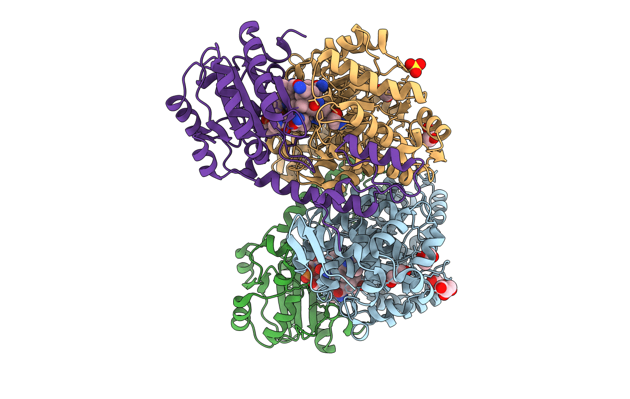
PDB ID:
3ABS
Keywords:
Title:
Crystal structure of ethanolamine ammonia-lyase from Escherichia coli complexed with adeninylpentylcobalamin and ethanolamine
Biological Source:
Source Organism(s):
Escherichia coli (Taxon ID: 83333)
Expression System(s):
Method Details:
Experimental Method:
Resolution:
2.25 Å
R-Value Free:
0.24
R-Value Work:
0.21
R-Value Observed:
0.21
Space Group:
P 63
