3A9F image
Deposition Date 2009-10-25
Release Date 2010-03-02
Last Version Date 2024-03-13
Entry Detail
PDB ID:
3A9F
Title:
Crystal structure of the C-terminal domain of cytochrome cz from Chlorobium tepidum
Biological Source:
Source Organism:
Host Organism:
Method Details:
Experimental Method:
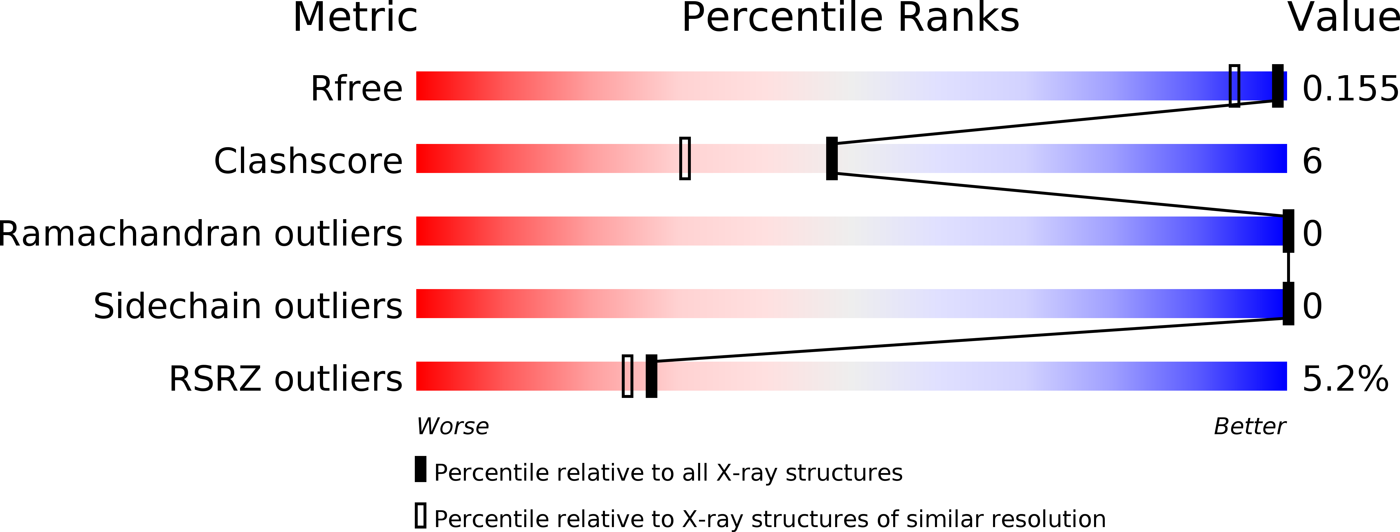
Resolution:
1.30 Å
R-Value Free:
0.15
R-Value Work:
0.13
R-Value Observed:
0.13
Space Group:
I 41
Macromolecular Entities
Polymer Type:polypeptide(L)
Molecule:Cytochrome c
Gene (Uniprot):pscC
Chain IDs:A, B
Chain Length:92
Number of Molecules:2
Biological Source:Chlorobaculum tepidum
Primary Citation
Crystal structure of the electron carrier domain of the reaction center cytochrome c(z) subunit from green photosynthetic bacterium Chlorobium tepidum
J.Mol.Biol. 397 1175 1187 (2010)
PMID: 20156447 DOI: 10.1016/j.jmb.2010.02.011

Abstact

In green sulfur photosynthetic bacteria, the cytochrome c(z) (cyt c(z)) subunit in the reaction center complex mediates electron transfer mainly from menaquinol/cytochrome c oxidoreductase to the special pair (P840) of the reaction center. The cyt c(z) subunit consists of an N-terminal transmembrane domain and a C-terminal soluble domain that binds a single heme group. The periplasmic soluble domain has been proposed to be highly mobile and to fluctuate between oxidoreductase and P840 during photosynthetic electron transfer. We have determined the crystal structure of the oxidized form of the C-terminal functional domain of the cyt c(z) subunit (C-cyt c(z)) from thermophilic green sulfur bacterium Chlorobium tepidum at 1.3-A resolution. The overall fold of C-cyt c(z) consists of four alpha-helices and is similar to that of class I cytochrome c proteins despite the low similarity in their amino acid sequences. The N-terminal structure of C-cyt c(z) supports the swinging mechanism previously proposed in relation with electron transfer, and the surface properties provide useful information on possible interaction sites with its electron transfer partners. Several characteristic features are observed for the heme environment: These include orientation of the axial ligands with respect to the heme plane, surface-exposed area of the heme, positions of water molecules, and hydrogen-bond network involving heme propionate groups. These structural features are essential for elucidating the mechanism for regulating the redox state of cyt c(z).

Legend

Protein

Chemical

Disease

Primary Citation of related structures