Deposition Date
2009-10-07
Release Date
2009-11-24
Last Version Date
2024-03-13
Entry Detail
PDB ID:
3A8Q
Keywords:
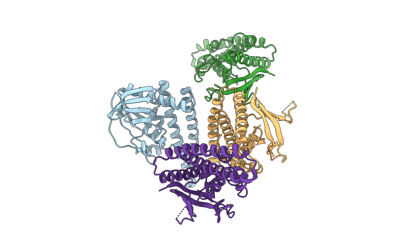
Title:
Low-resolution crystal structure of the Tiam2 PHCCEx domain
Biological Source:
Source Organism(s):
Mus musculus (Taxon ID: 10090)
Expression System(s):
Method Details:
Experimental Method:
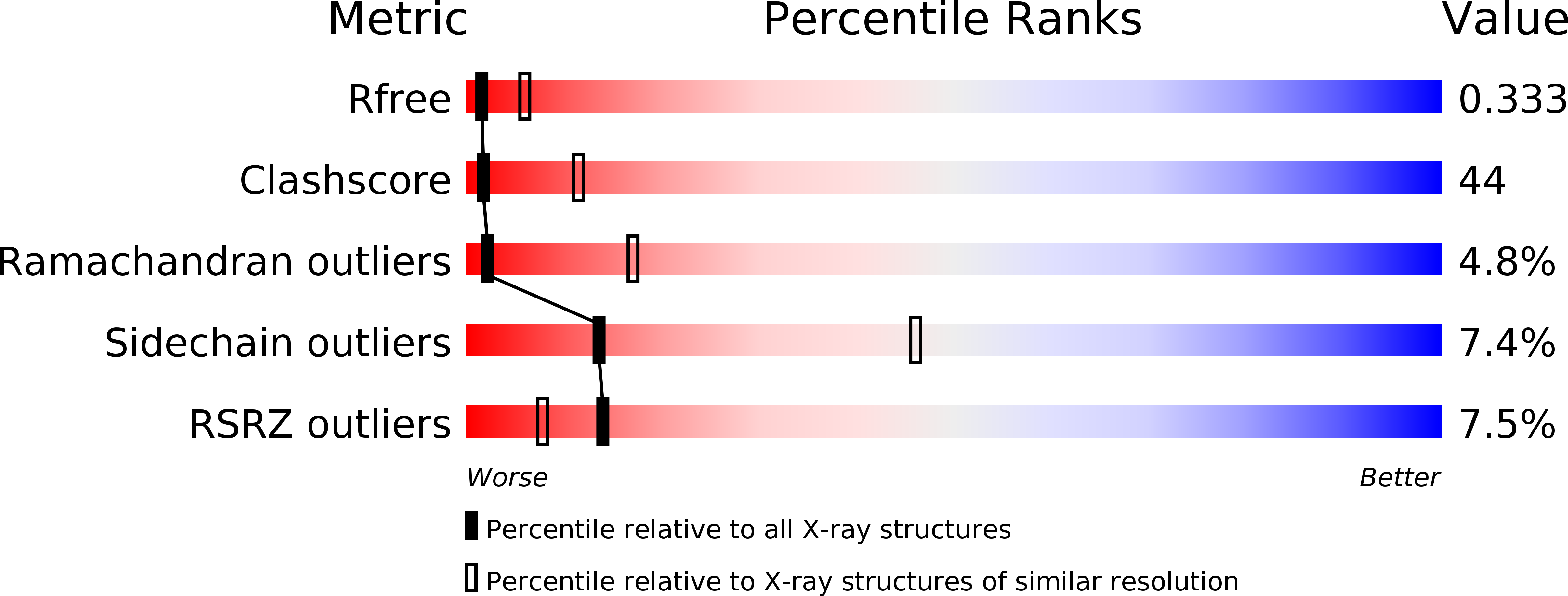
Resolution:
3.20 Å
R-Value Free:
0.33
R-Value Work:
0.28
R-Value Observed:
0.28
Space Group:
P 43 21 2