Deposition Date
2009-10-06
Release Date
2010-04-14
Last Version Date
2024-10-23
Entry Detail
PDB ID:
3A8L
Keywords:
Title:
Crystal structure of photo-activation state of Nitrile Hydratase mutant S113A
Biological Source:
Source Organism(s):
Rhodococcus erythropolis (Taxon ID: 1833)
Expression System(s):
Method Details:
Experimental Method:
Resolution:
1.63 Å
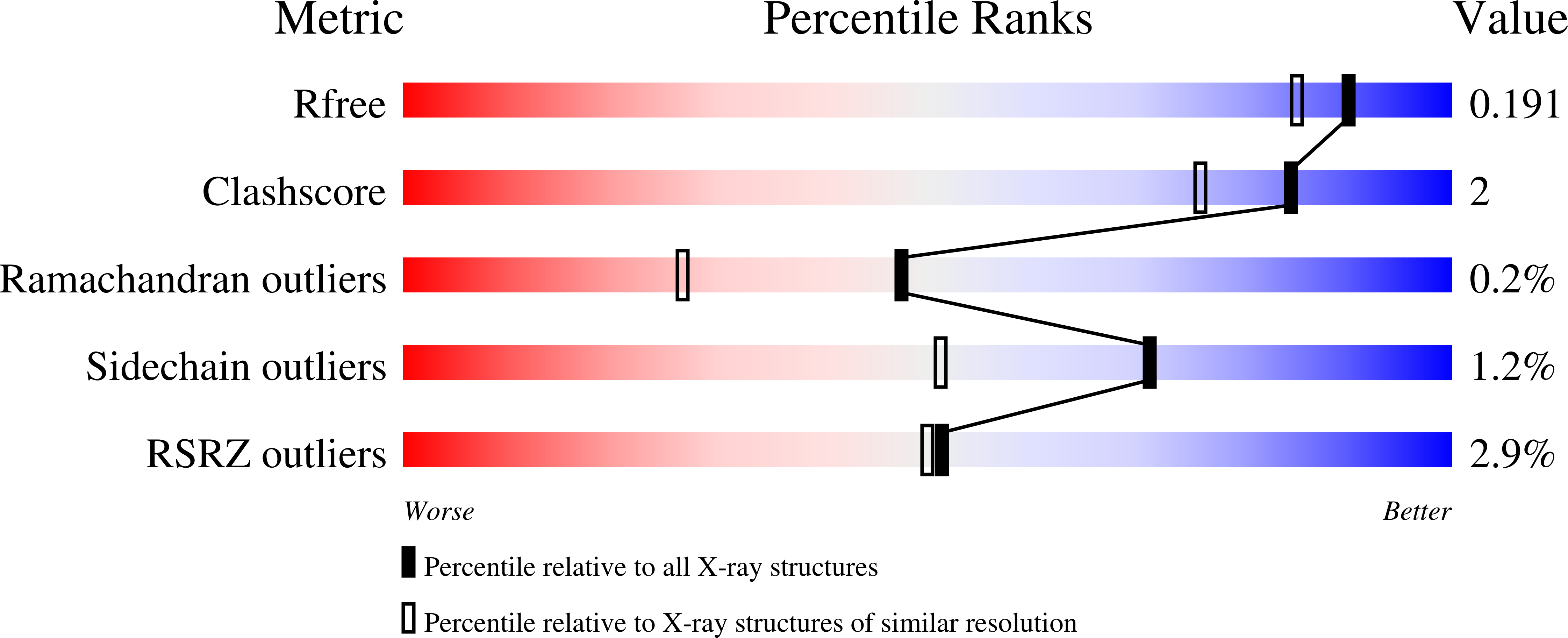
R-Value Free:
0.19
R-Value Work:
0.16
R-Value Observed:
0.16
Space Group:
C 1 2 1