3A7N image
Deposition Date 2009-09-29
Release Date 2010-08-11
Last Version Date 2023-11-01
Entry Detail
PDB ID:
3A7N
Keywords:
Title:
Crystal structure of uracil-DNA glycosylase from Mycobacterium tuberculosis
Biological Source:
Source Organism:
Host Organism:
Method Details:
Experimental Method:
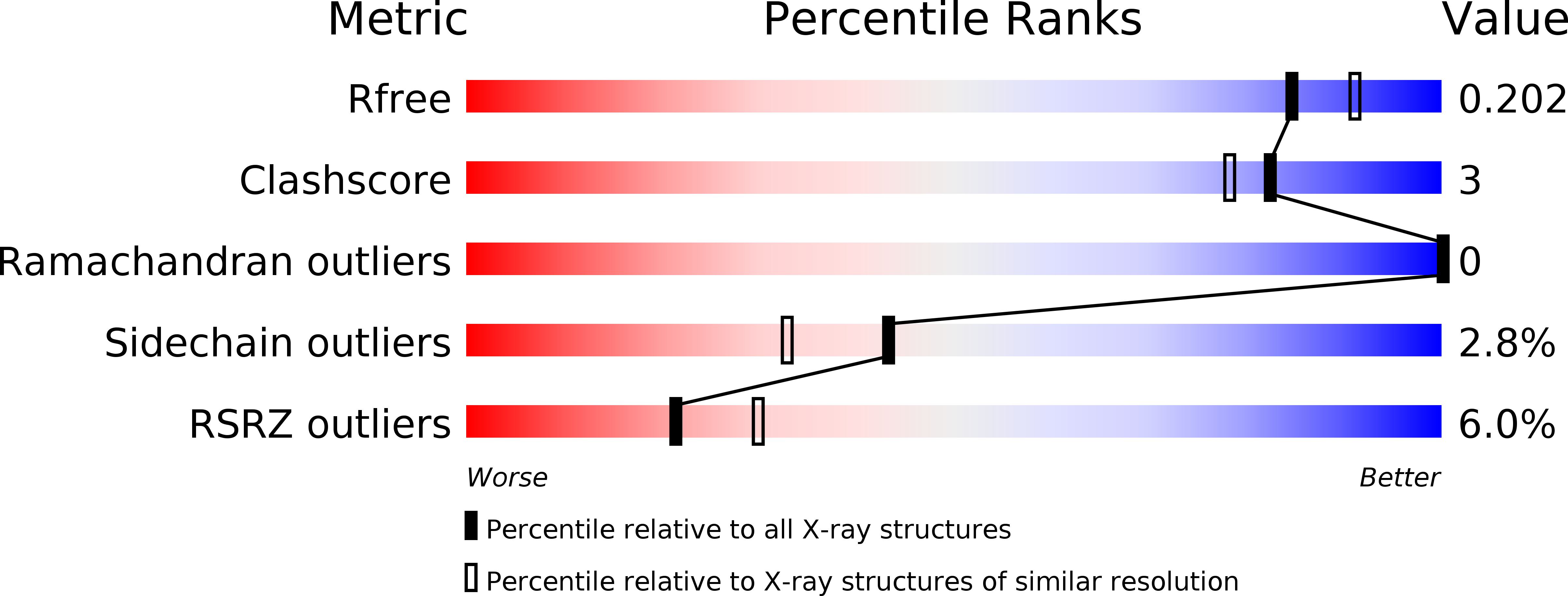
Resolution:
1.95 Å
R-Value Free:
0.19
R-Value Work:
0.18
R-Value Observed:
0.18
Space Group:
P 21 21 21
Macromolecular Entities
Polymer Type:polypeptide(L)
Molecule:Uracil-DNA glycosylase
Chain IDs:A
Chain Length:238
Number of Molecules:1
Biological Source:Mycobacterium tuberculosis H37Rv
Ligand Molecules
Primary Citation
Structure of uracil-DNA glycosylase from Mycobacterium tuberculosis: insights into interactions with ligands
Acta Crystallogr.,Sect.F 66 887 892 (2010)
PMID: 20693660 DOI: 10.1107/S1744309110023043

Abstact

Uracil N-glycosylase (Ung) is the most thoroughly studied of the group of uracil DNA-glycosylase (UDG) enzymes that catalyse the first step in the uracil excision-repair pathway. The overall structure of the enzyme from Mycobacterium tuberculosis is essentially the same as that of the enzyme from other sources. However, differences exist in the N- and C-terminal stretches and some catalytic loops. Comparison with appropriate structures indicate that the two-domain enzyme closes slightly when binding to DNA, while it opens slightly when binding to the proteinaceous inhibitor Ugi. The structural changes in the catalytic loops on complexation reflect the special features of their structure in the mycobacterial protein. A comparative analysis of available sequences of the enzyme from different sources indicates high conservation of amino-acid residues in the catalytic loops. The uracil-binding pocket in the structure is occupied by a citrate ion. The interactions of the citrate ion with the protein mimic those of uracil, in addition to providing insights into other possible interactions that inhibitors could be involved in.

Legend

Protein

Chemical

Disease

Primary Citation of related structures