Abstact
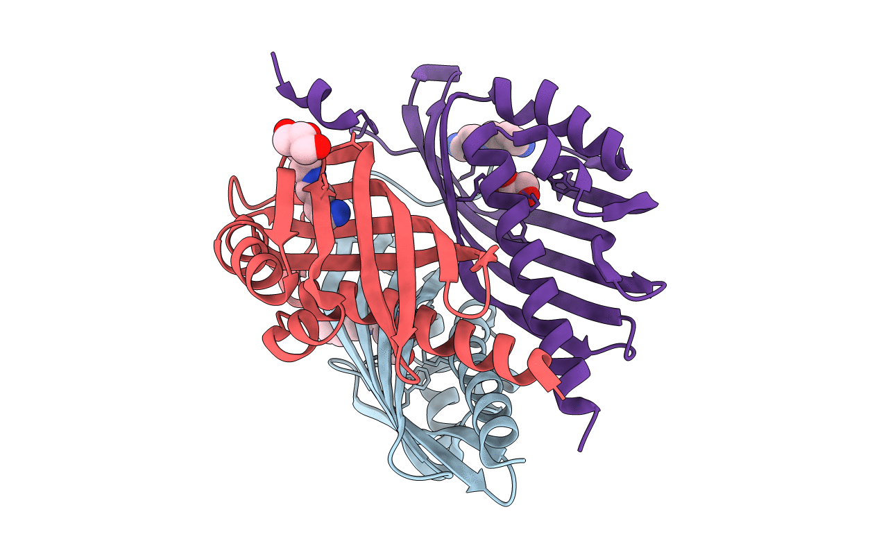
LinA from Sphingobium japonicum UT26 catalyzes two steps of dehydrochlorination from γ hexachlorocyclohexane (HCH) to 1,3,4,6-tetrachloro-1,4-cyclohexadiene via γ-pentachlorocyclohexene. We determined the crystal structure of LinA at 2.25 Å by single anomalous dispersion. LinA exists as a homotrimer, and each protomer forms a cone-shaped α+β barrel fold. The C-terminal region of LinA is extended to the neighboring subunit, unlike that of scytalone dehydratase from Magnaporthe grisea, which is one of the most structurally similar proteins identified by the DALI server. The structure we obtained in this study is in open form, in which γ-HCH can enter the active site. There is a hydrophobic cavity inside the barrel fold, and the active site is largely surrounded by the side chains of K20, L21, V24, D25, W42, L64, F68, C71, H73, V94, L96, I109, F113, and R129. H73 was considered to function as a base that abstracts the proton of γ-HCH through its interaction with D25. Docking simulations with γ-HCH and γ-pentachlorocyclohexene suggest that 11 residues (K20, I44, L64, V94, L96, I109, A111, F113, A131, C132, and T133) are involved in the binding of these compounds and support the degradation mechanism.