
Deposition Date
2009-09-10
Release Date
2010-05-26
Last Version Date
2023-11-01
Entry Detail
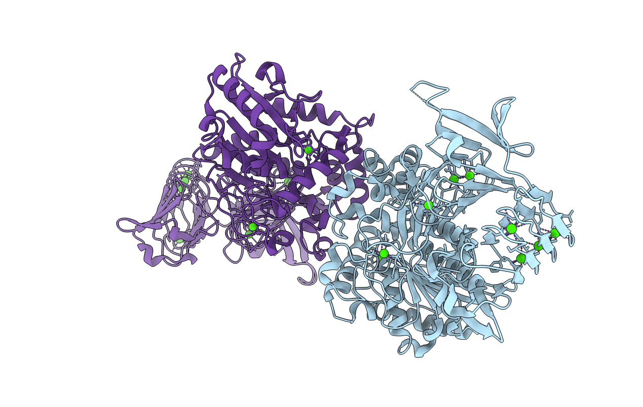
PDB ID:
3A6Z
Keywords:
Title:
Crystal structure of Pseudomonas sp. MIS38 lipase (PML) in the open conformation following dialysis against Ca-free buffer
Biological Source:
Source Organism(s):
Pseudomonas (Taxon ID: 91465)
Expression System(s):
Method Details:
Experimental Method:
Resolution:
2.15 Å
R-Value Free:
0.21
R-Value Work:
0.18
R-Value Observed:
0.18
Space Group:
P 65 2 2
